 |
|
You are here: Medical Histology>Main Web>AtlasContents>FemaleReproductiveSystemAtlas16 (20 Jun 2015, LorenEvey)Edit Attach
Chapter Sixteen: Female Reproductive System


- Introduction
- Ovary
- Female Duct System
- Uterus
- Uterine Cervix
- Vagina
- Mammary Gland
- Placenta
- Chapter Sixteen Review
- Comments
Introduction
The female reproductive system consists of the paired gonads, the ovaries, as well as a system of auxiliary structures, the accessory sexual organs. The latter includes the uterine tubes, uterus, and vagina. Although not a genital organ, the mammary glands are an important adjunct to this system and are included herein. It is important to note that virtually every part of this system is dependent, both structurally and functionally, on the levels of certain circulating hormones. Further, these levels change over the life of the female (menarche, pregnancy, and menopause) as well as during the reproductive years when they fluctuate with continuous regularity thereby defining the menstrual cycle. As the changing hormone levels elicit various functional activities, changes are seen in morphology. Prior to learning the microarchitecture of the Female Reproductive Tract, use the table below to review some of the gross anatomy of these tissues:| Structure | Image |
|---|---|
| Cross Section of the Ovary | |
| Arrangement of the Uterus, Ovary, and Uterine Tubes | |
| Gross Anatomical Location of the Uterus in the Pelvic Cavity | |
| Arrangement of the Uterus, Cervix, and Vagina | |
| The Mammary Gland | |
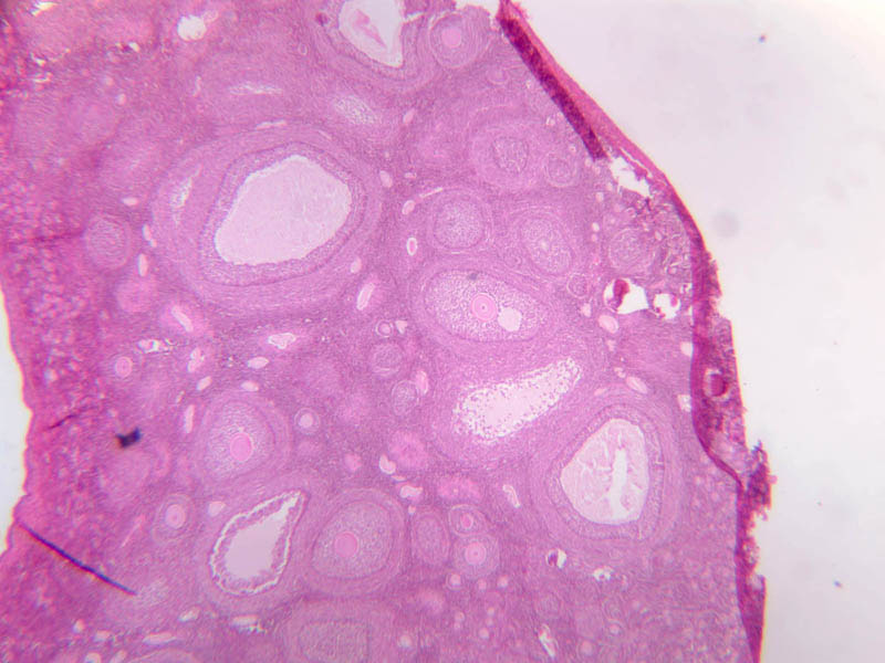
Ovary
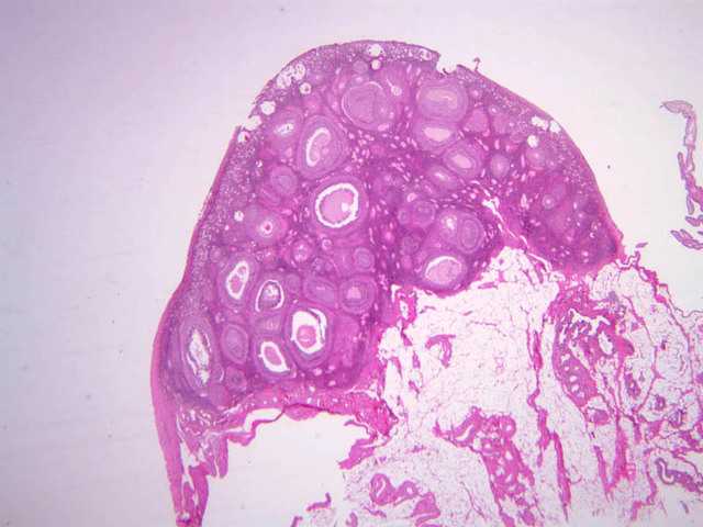
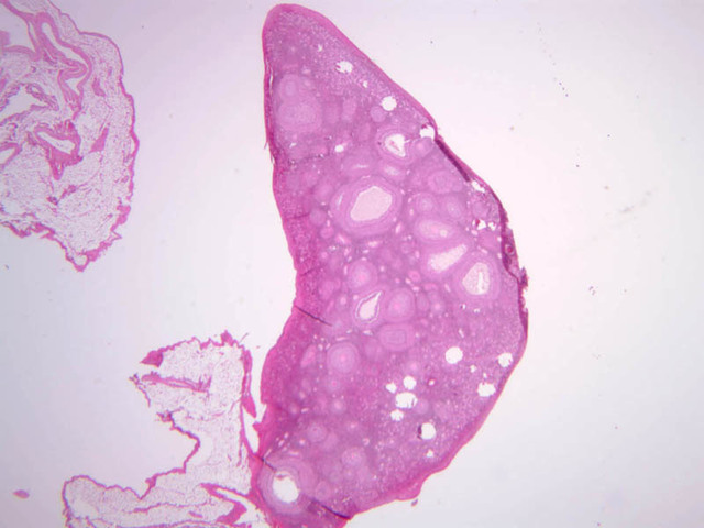
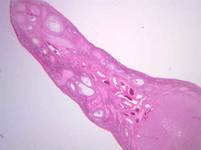
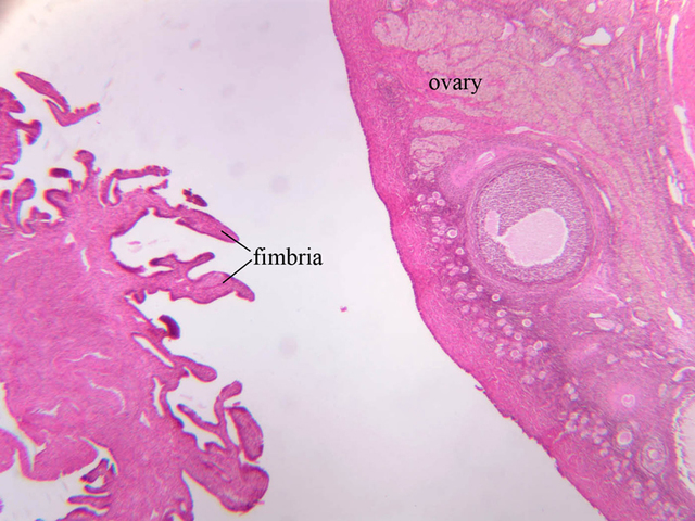
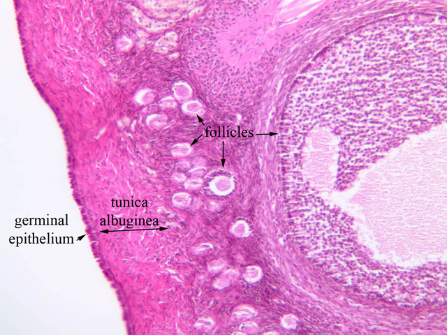
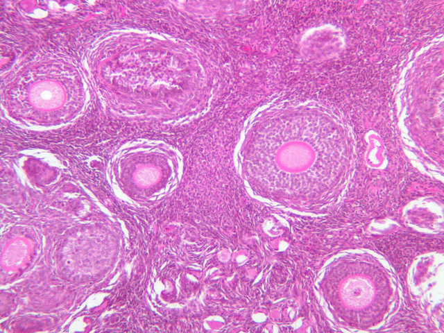
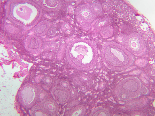
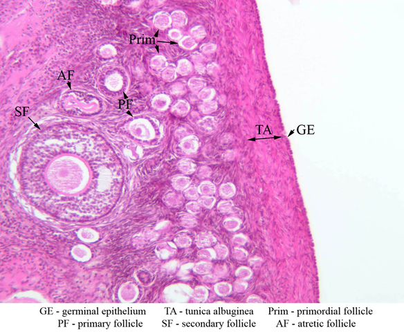
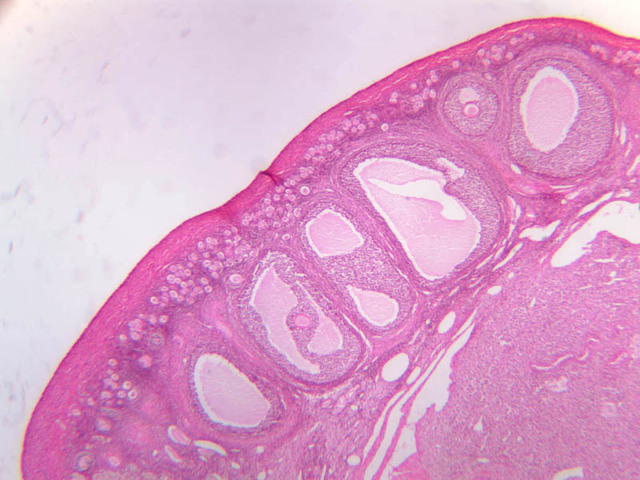
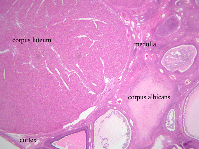
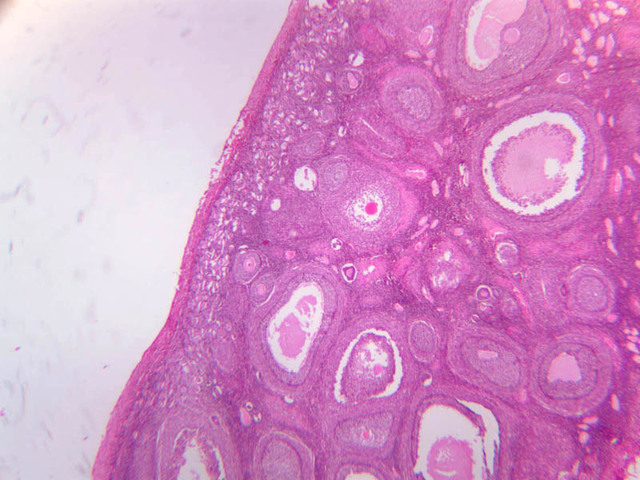
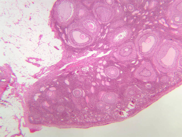
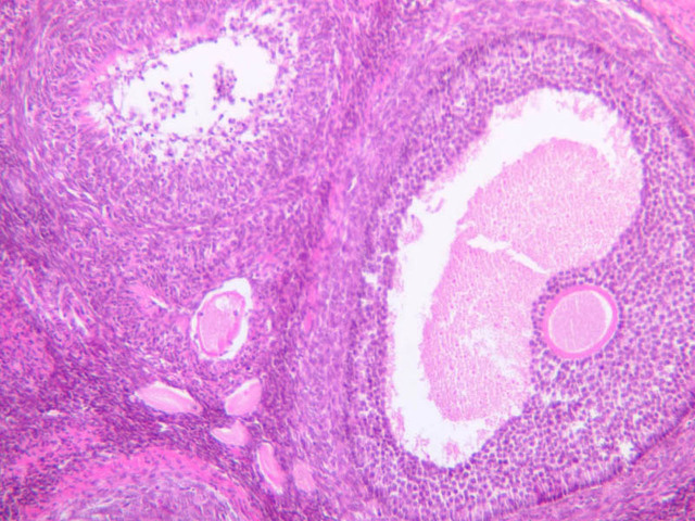
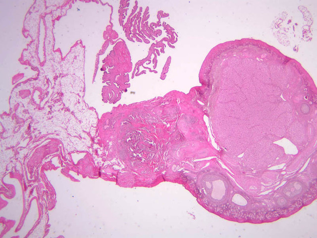
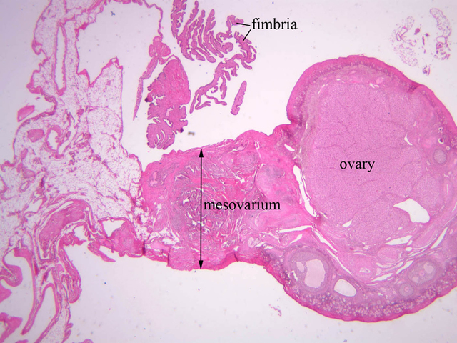
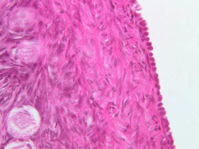
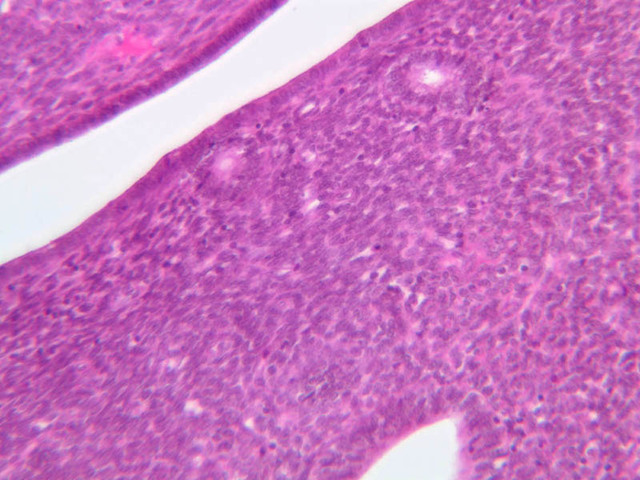
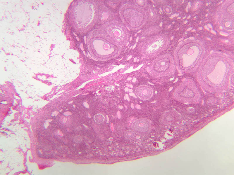
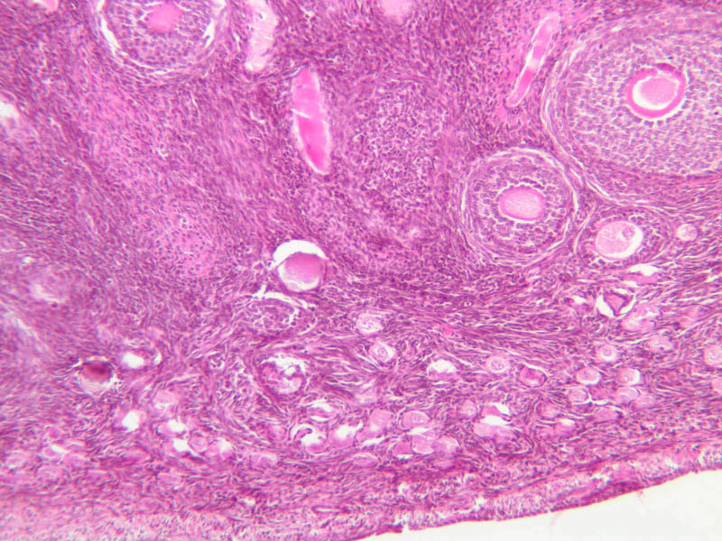
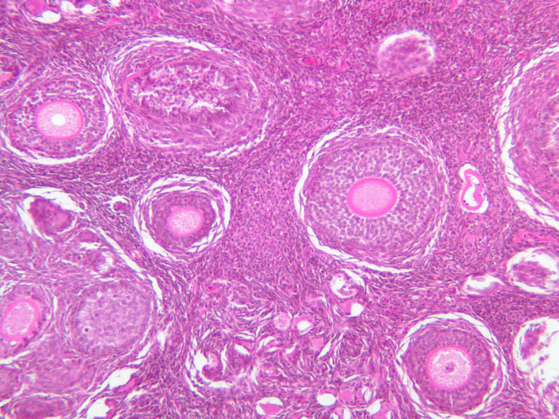
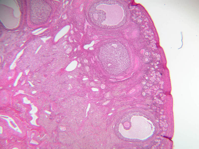
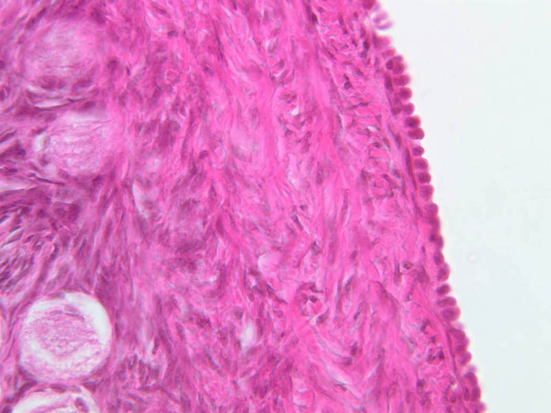
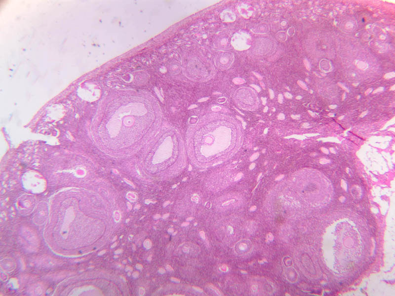
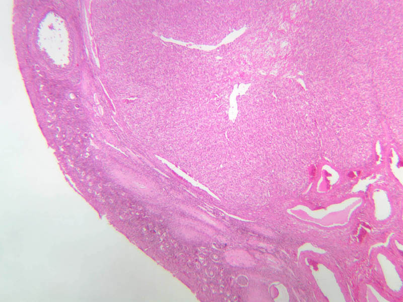
The ovary is the site where ova develop and hormones are produced which in turn have direct action in maintaining the cycle. Its structure varies both with the age of the female as well as throughout the cycle. The ovary is a somewhat oval and slightly flattened organ situated near the fimbriated end of the uterine tube (B-94, monkey ovary, H&E [1x, 1x]; B-95, monkey ovary, H&E [1x-labeled, 1x]; B-96, monkey ovary and tube, H&E [1x]; B-54, H&E [1x, 1x]). Its exposed surface is covered by a mesothelial sheet of squamous or cuboidal epithelium. Just deep to this is an ill defined area of dense fibrous connective tissue referred to as the tunica albuginea (B-96 [2.5x-labeled, 10x-labeled, 20x, 40x]). The ovary has two major regions – the cortex and the medulla. The cortex is the broad peripheral area containing follicles in various stages of development, whereas the medulla is the more central area displaying profiles of large blood vessels. These blood vessels gain entrance via the mesovarium. The division between the cortex and medulla is indistinct. In the cortex, stromal cells occupy the areas between the follicles. These cells are closely packed, fusiform (spindle shaped), and have the potential to differentiate into a specialized component of the maturing follicle (theca folliculi). With each cycle several follicles begin the process of follicular growth and maturation. The initial morphological event in this process is the transformation of primordial follicles to primary follicles. Normally only one follicle achieves the mature state and ovulates per cycle. The remainder cease growing and degenerate at various points in the maturation process. After ovulation, at mid-cycle, the ruptured follicle persists for a short while as the functionally important corpus luteum.Ovary Image Gallery


Ovary Table of Identifications
| Row | Structure | Abbreviation | Optimal Stain | Representative Section | Note |
|---|---|---|---|---|---|
| 1 | Fimbria | (none) | H&E | |
|
| 2 | Mesovarium | (none) | H&E | |
|
| 3 | Ovary | (none) | H&E | |
|
| 4 | Corpus Luteum | (none) | H&E | |
|
| 5 | Medulla (Ovarian) | (none) | H&E | |
|
| 6 | Cortex (Ovarian) | (none) | H&E | |
|
| 7 | Corpus Albicans | (none) | H&E | |
|
| 8 | Follicles | (none) | H&E | |
|
| 9 | Tunica Albuginea | (none) | H&E | |
|
| 10 | Germinal Epithelium | (none) | H&E | |
Top of page
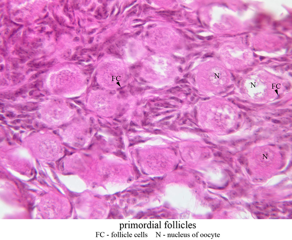
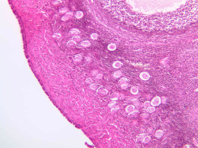
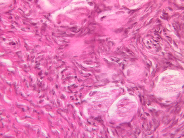
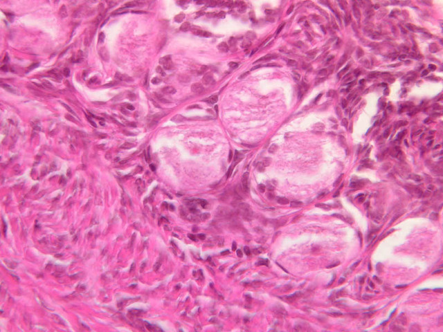
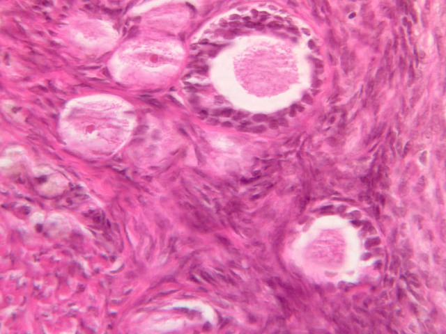
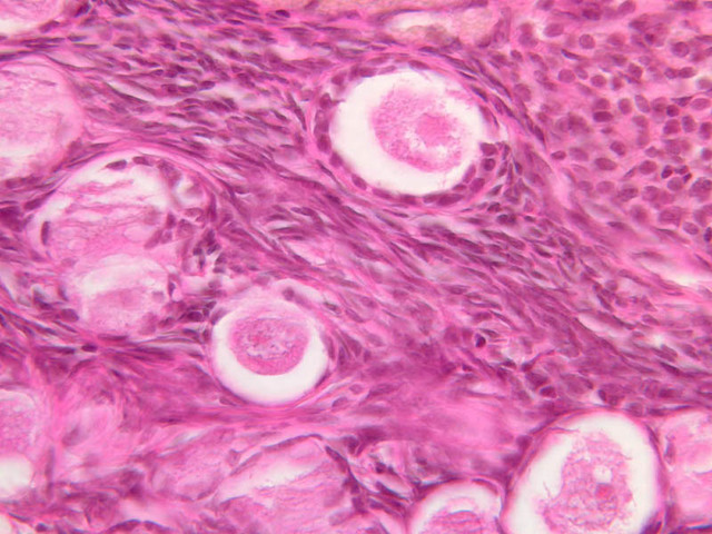
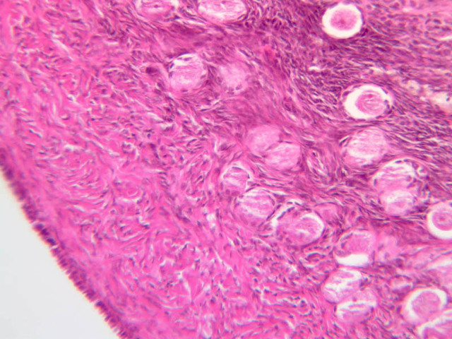
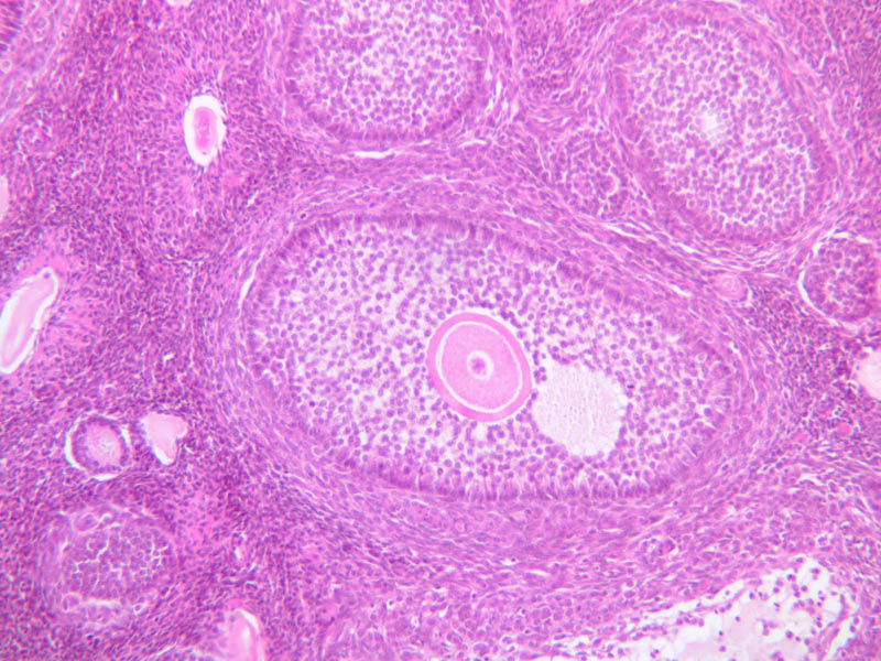
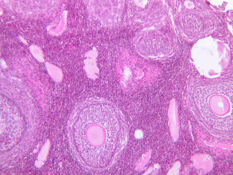
Primordial Follicle
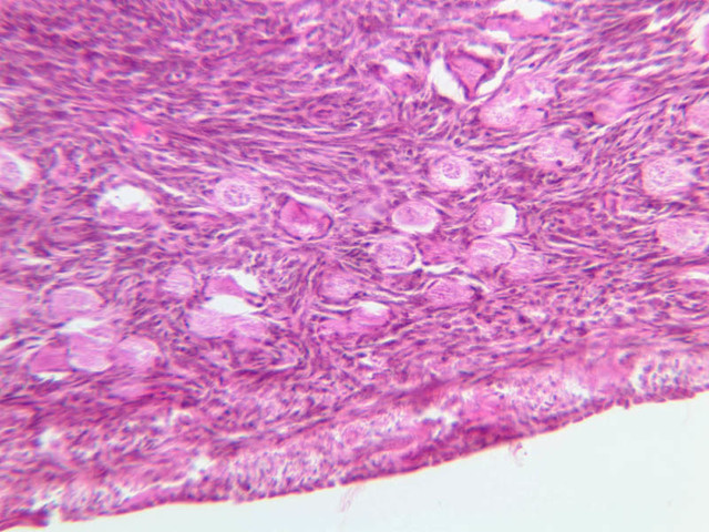
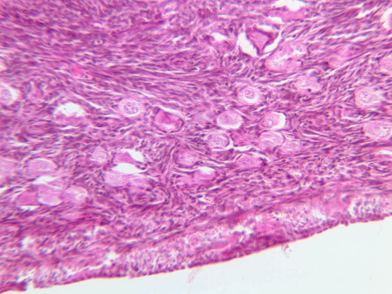
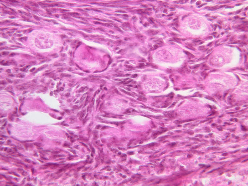
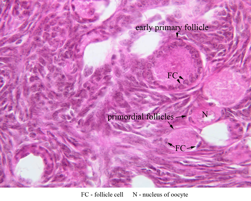
Primordial follicles are located in periphery of cortex. They contain a large round oocyte surrounded by a single layer of flattened follicular cells. The follicles occupying this category are inactive. They are the population from which follicular growth begins. Primordial Follicles [Inactive] (slide B-94 [2.5x, 10x, 20x, 40x] [10x, 20x, 40x-labeled] [10x, 20x, 40x]; B-96 [2.5x, 10x, 20x, 40x, 40x, 40x, 40x]).Primordial Follicle Image Gallery


Primordial Follicle Table of Identifications
| Row | Structure | Abbreviation | Optimal Stain | Representative Section | Note |
|---|---|---|---|---|---|
| 1 | Primordial Follicle | (none) | H&E | |
|
| 2 | Follicle Cells | FC | H&E | |
|
| 3 | Nucleus of Oocyte | N | H&E | |
Top of page
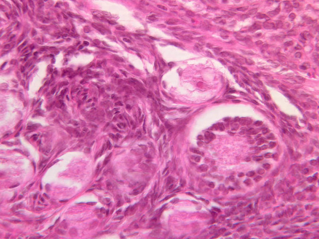
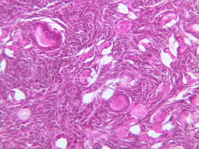
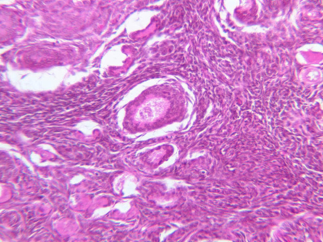
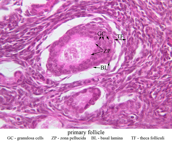
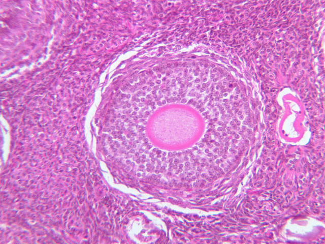
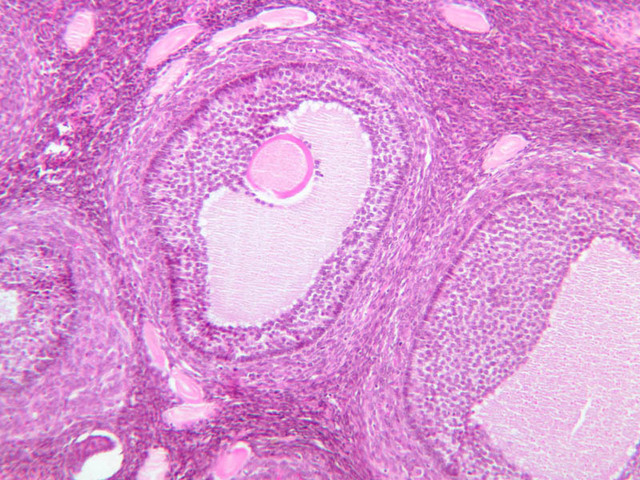
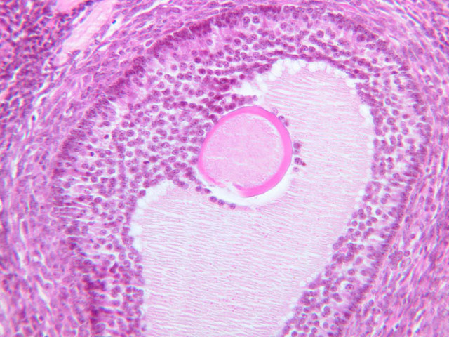
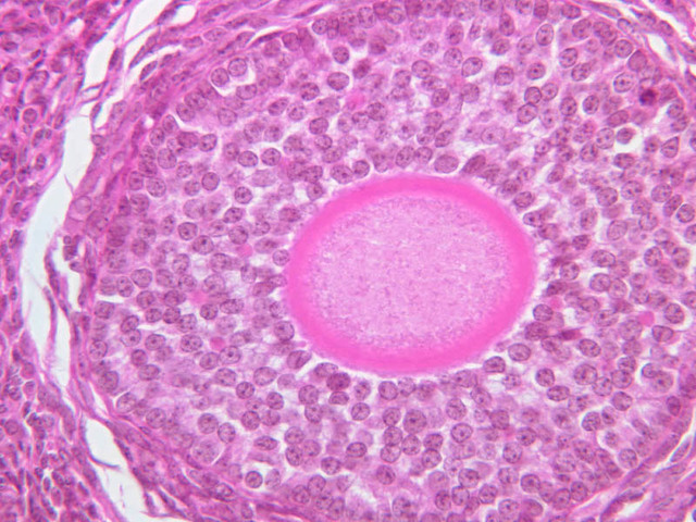
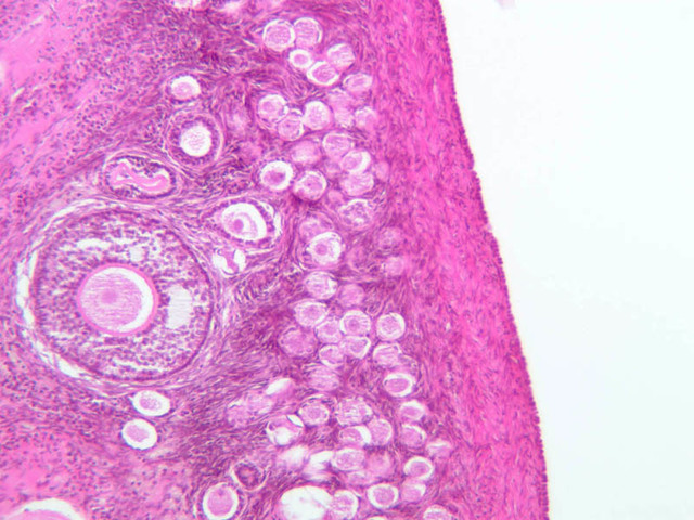
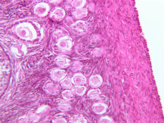
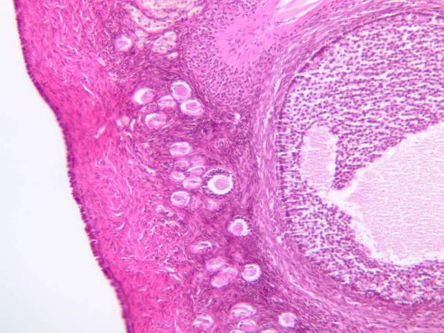
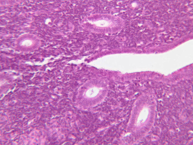
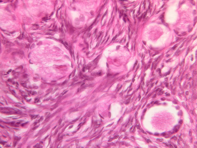
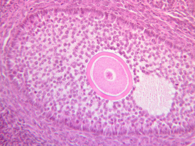
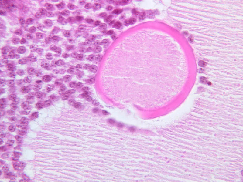
Primary Follicle
- Early primary follicles are characterized by:
- Oocyte becomes enlarged.
- Oocyte nucleus is large, eccentrically placed and vesicular.
- The follicular cells have increased in size and are cuboidal.
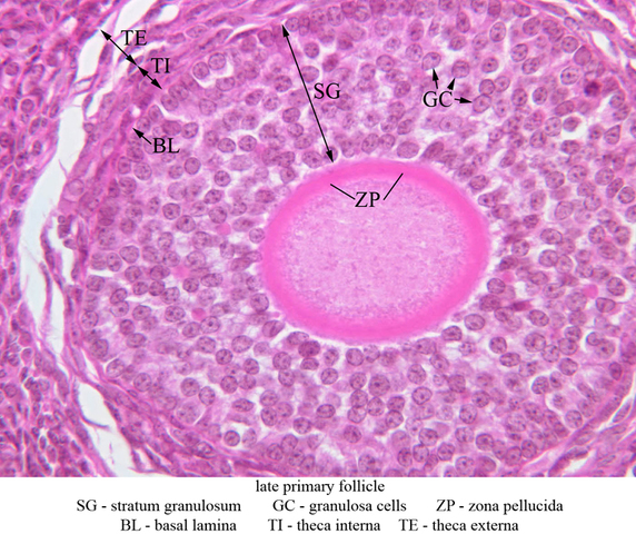
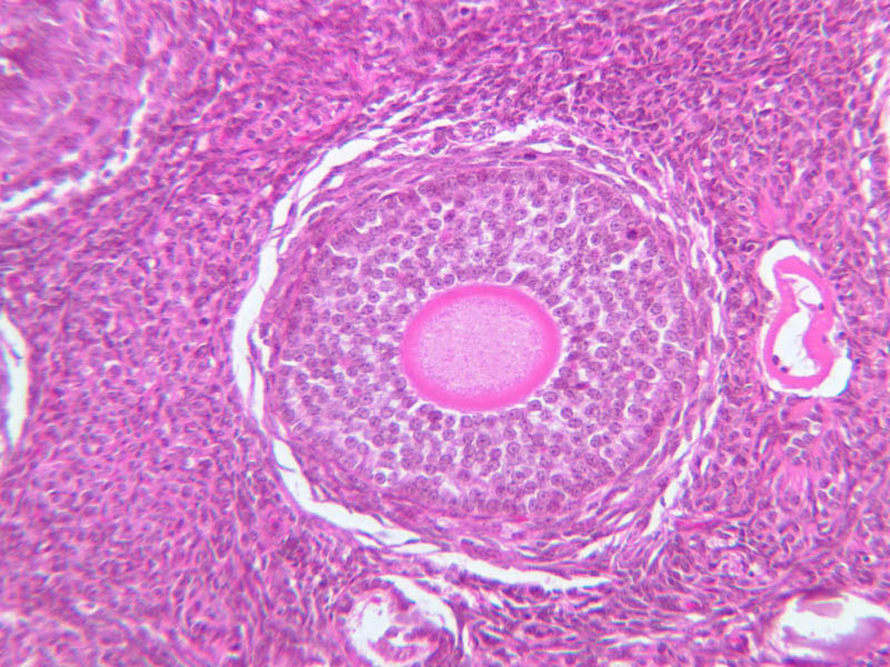
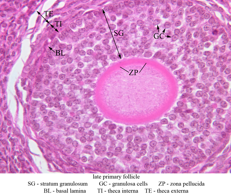
- Later primary follicles are characterized by:
- Follicular cells have proliferated so that the oocyte is surrounded by multiple layers of follicular cells, now referred to as granulosa cells.
- Eosinophilic zona pellucida appears and separates oocyte from the granulosa cells.
- Cortical stromal cells become arranged in a compressed layer at the outer periphery of the granulosa cells, thus forming the outer wall of the follicle. This layer is referred to as the theca folliculi.
Primary Follicle Image Gallery


Primary Follicle Table of Identifications
| Row | Structure | Abbreviation | Optimal Stain | Representative Section | Note |
|---|---|---|---|---|---|
| 1 | Early Primary Follicle | (none) | H&E | |
|
| 2 | Primordial Follicles | (none) | H&E | |
|
| 3 | Follicle Cell | FC | H&E | |
|
| 4 | Nucleus of Oocyte | N | H&E | |
|
| 5 | Granulosa Cells | GC | H&E | |
|
| 6 | Zona Pellucida | ZP | H&E | |
|
| 7 | Basal Lamina | BL | H&E | |
|
| 8 | Theca Folliculi | TF | H&E | |
|
| 9 | Late Primary Follicle | (none) | H&E | |
|
| 10 | Stratum Granulosum | GC | H&E | |
|
| 11 | Theca Interna | TI | H&E | |
|
| 12 | Theca Externa | TE | H&E | |
Top of page
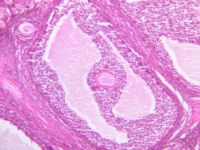
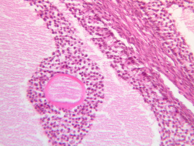
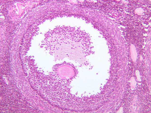
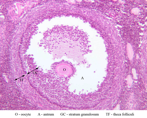
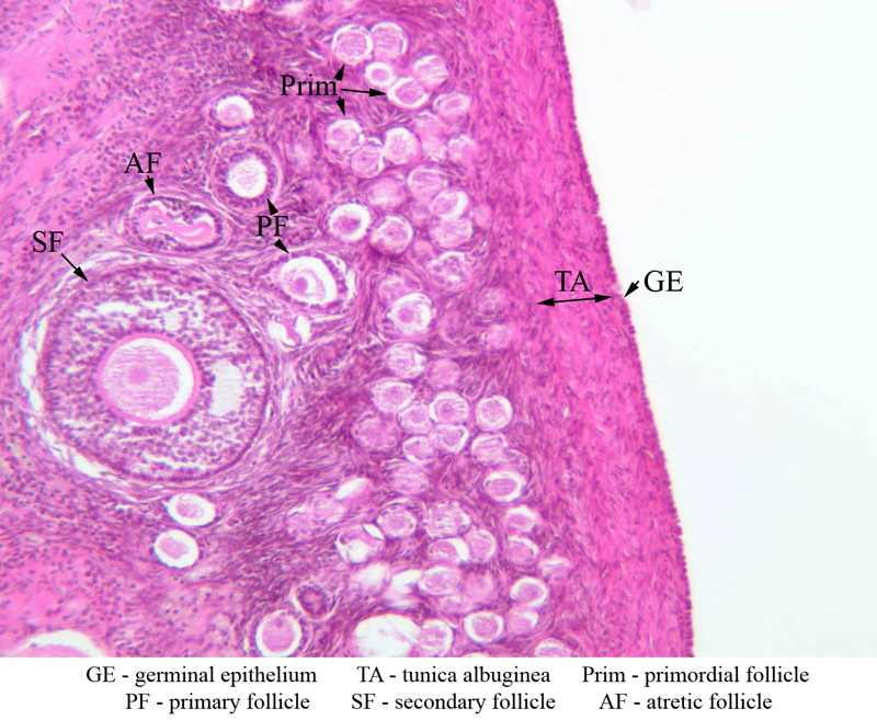
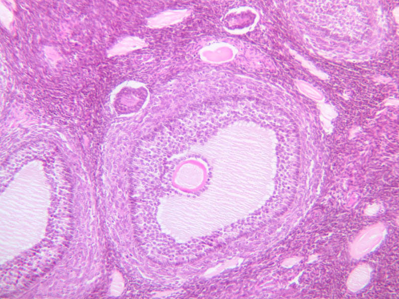
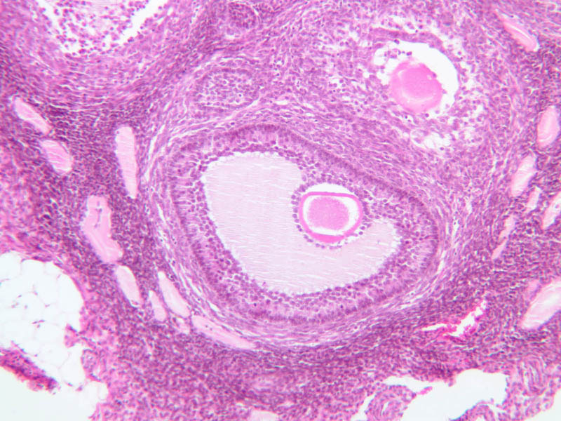
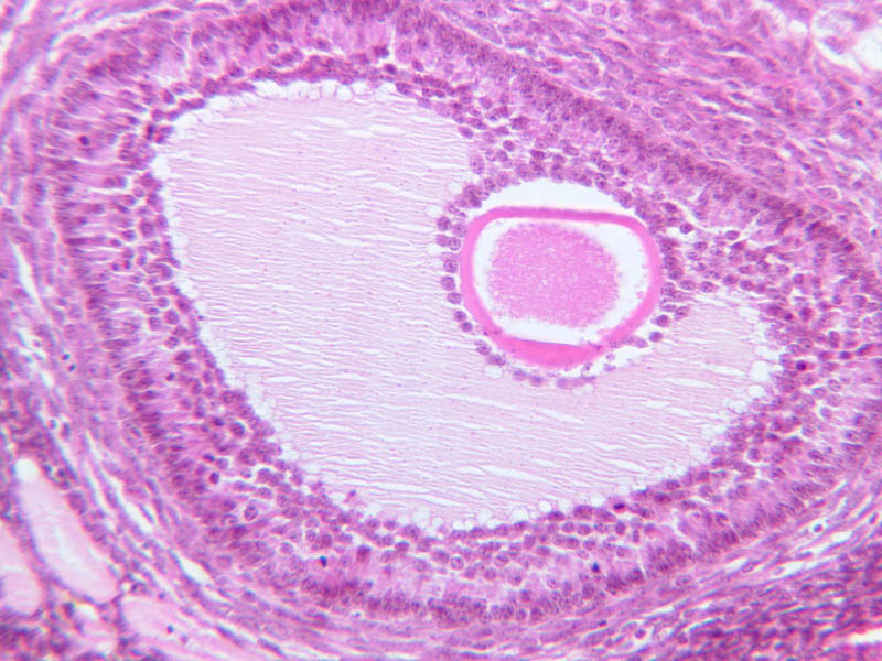
Secondary Follicle
- Early (vesicular stage)
- With continued granulosa cell proliferation, spaces filled with fluid (liquor folliculi) appear within this layer.
- Later (antral stage)
- Continued follicular enlargement occurs and the irregular spaces within the granulosa layer merge to form a single crescentric cavity, the antrum.
- Those granulosa cells immediately surrounding the zona pellucida become columnar and are referred to as the corona radiata.
- The theca folliculi begins to segregate itself into two distinct layers.
Secondary Follicle Image Gallery


Secondary Follicle Table of Identifications
| Row | Structure | Abbreviation | Optimal Stain | Representative Section | Note |
|---|---|---|---|---|---|
| 1 | Oocyte | O | H&E | |
|
| 2 | Antrum | A | H&E | |
|
| 3 | Stratum Granulosum | GC | H&E | |
|
| 4 | Theca Folliculi | TF | H&E | |
|
| 5 | Germinal Epithelium | GE | H&E | |
|
| 6 | Tunica Albuginea | TA | H&E | |
|
| 7 | Primordial Follicle | Prim | H&E | |
|
| 8 | Primary Follicle | PF | H&E | |
|
| 9 | Secondary Follicle | SF | H&E | |
|
| 10 | Atretic Follicle | AF | H&E | |
Top of page
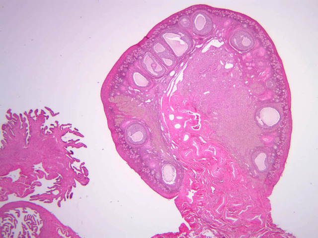
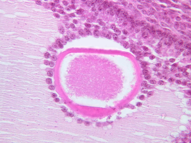
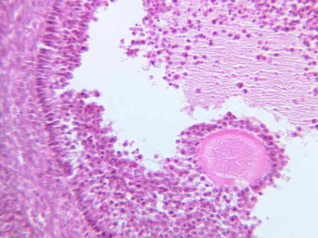
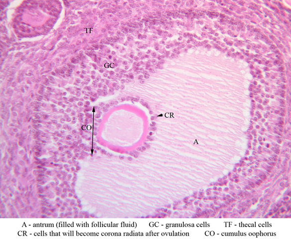
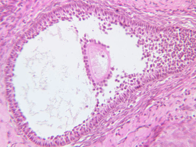
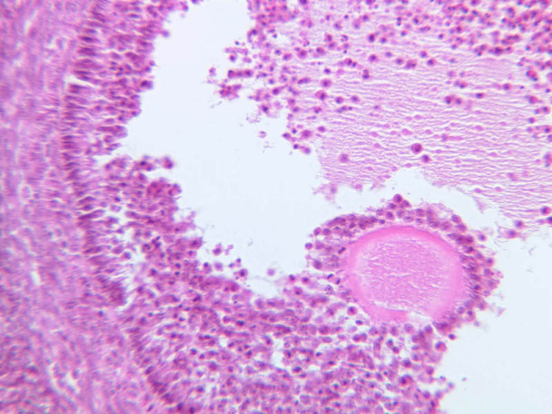
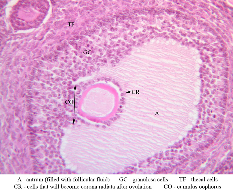
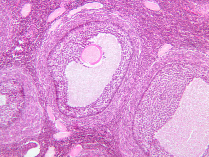
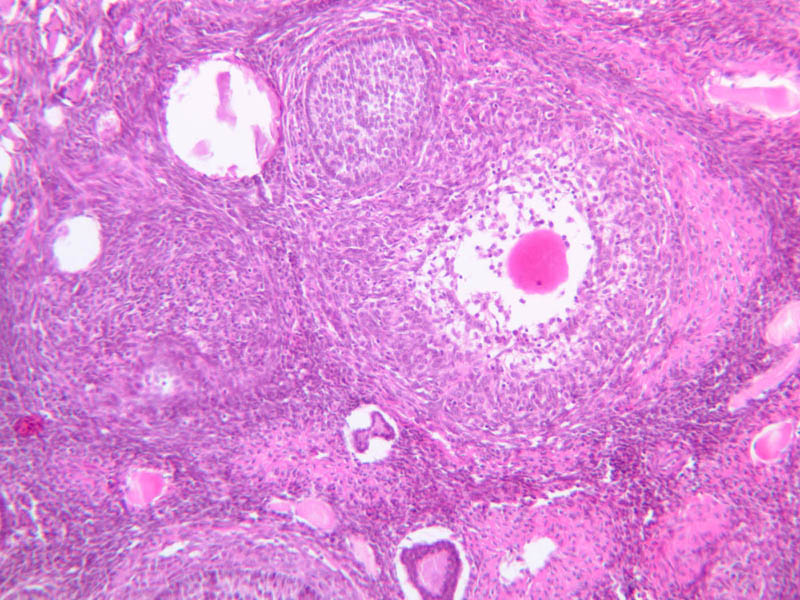
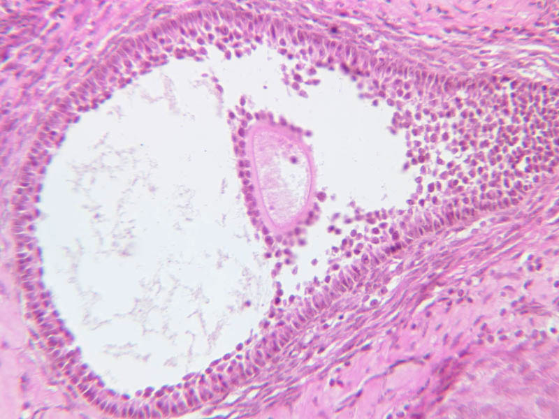
Mature (Graafian) Follicle
At maximum size, follicles will bulge from the ovarian surface and occupy the full thickness of the cortex. The oocyte is displaced to one side of the antrum where it is surrounded by a thickening of granulosa cells. This stalk-like thickening is referred to as the cumulus oophorus. A distinct basal lamina separates the granulosa cells from the theca. The theca has segregated itself into the theca interna and the theca externa. The theca interna is adjacent to the basal lamina, highly vascular and consists of secretory cells. The theca externa is connective tissue that merges with the adjacent stroma. Mature (Graafian) Follicle (slide B-94 [2.5x, 10x, 20x-labeled] [10x, 20x, 40x] [10x, 20x, 40x])Mature Follicle Image Gallery


Mature Follicle Table of Identifications
| Row | Structure | Abbreviation | Optimal Stain | Representative Section | Note |
|---|---|---|---|---|---|
| 1 | Antrum | A | H&E | |
|
| 2 | Granulosa Cells | GC | H&E | |
|
| 3 | Thecal Cells | TF | H&E | |
|
| 4 | Corona Radiata (future) | CR | H&E | |
|
| 5 | Cumulus Oophorus | CO | H&E | |
Top of page
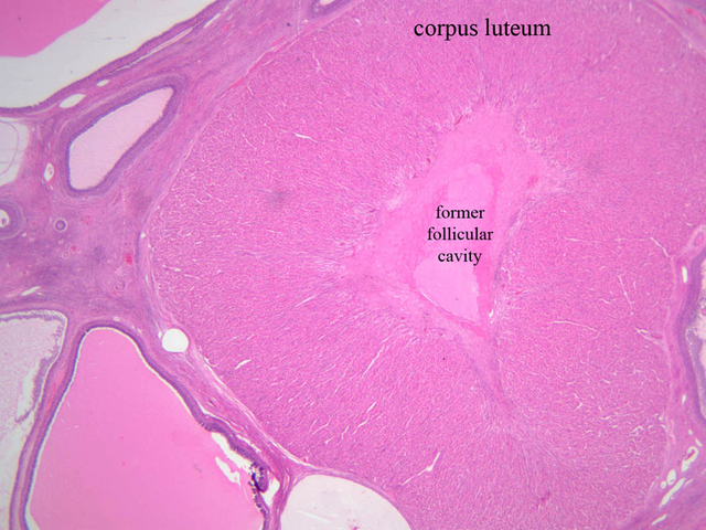
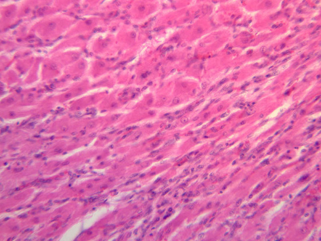
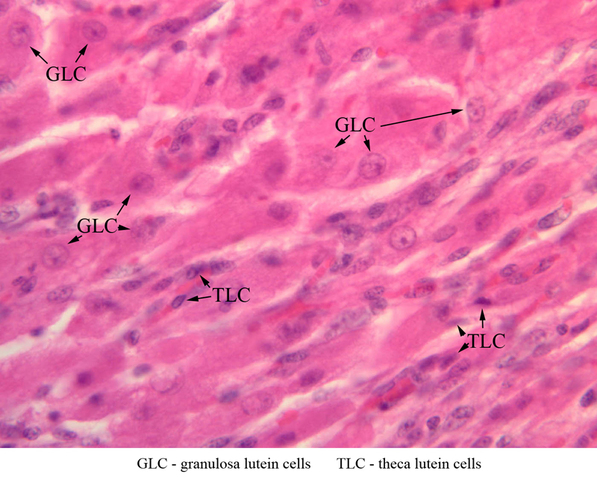
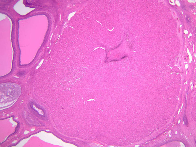
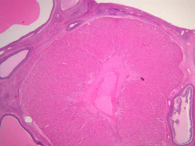
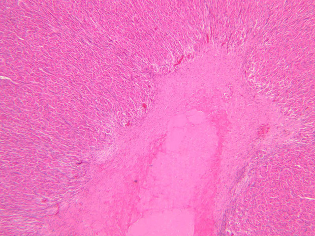
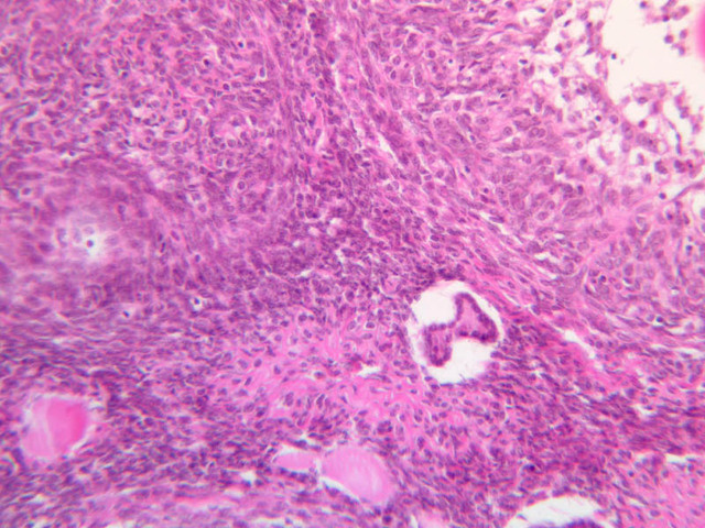
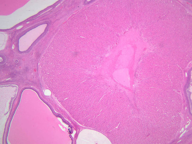
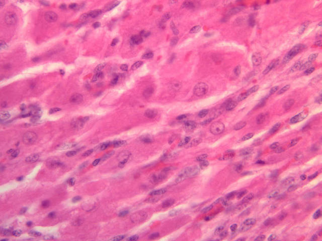
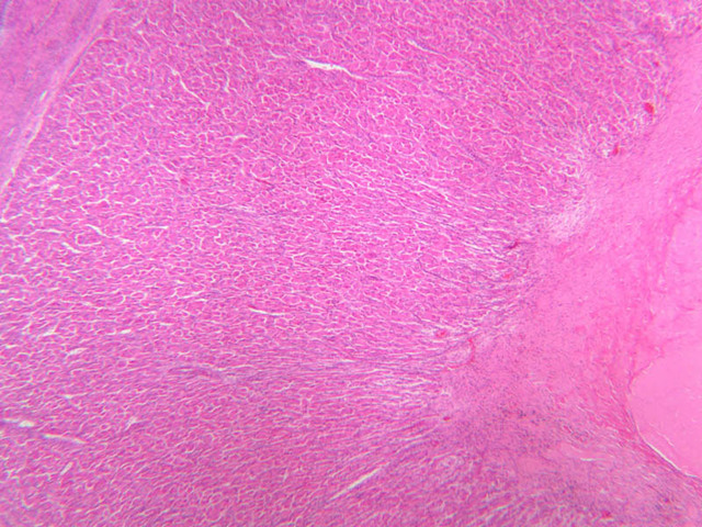
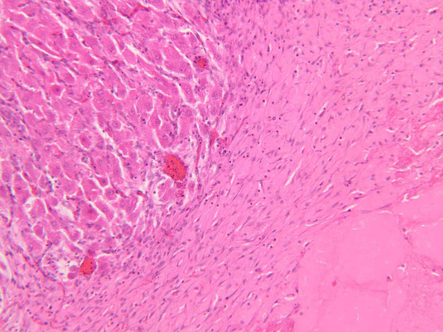
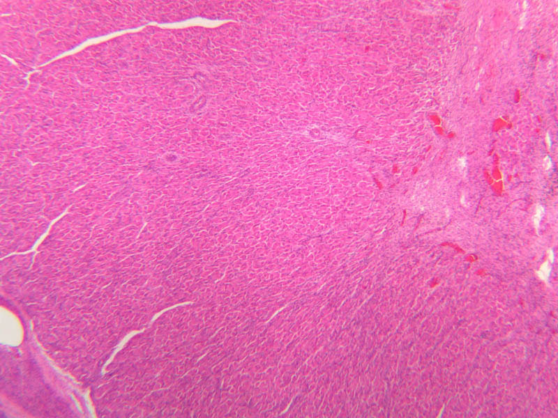
Corpus Luteum
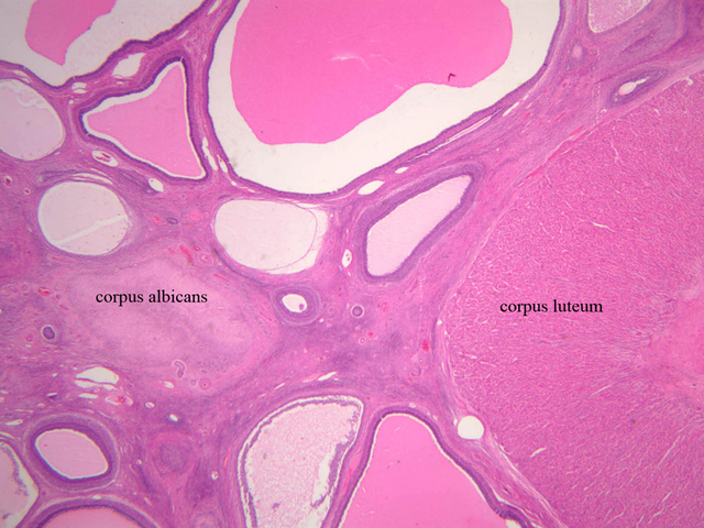
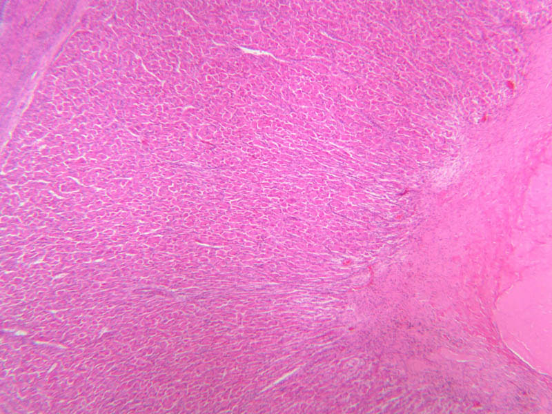
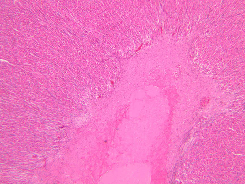
After rupture of the Graafian follicle, the walls collapse and become folded. Thus, strands of tissue from the theca interna penetrate, to some extent, the mass of granulosa cells. Bleeding from the theca interna may form a blood clot in the antrum. The majority of the cells of the corpus luteum are derived from the granulosa cells. Both the granulosa cells and the cells of the theca interna enlarge and accumulate lipid, thus becoming lutein cells. There are two types of lutein cells - granulosa lutein cells that have a large, vesicular nucleus and vacuolate cytoplasm, and theca lutein cells that are distinguished by smaller darker nuclei. Corpus Luteum (slide B-54, human ovary, H&E [1x-labeled, 2.5x, 10x, 20x, 40x-labeled] [1x-labeled, 1x, 2.5x] [1x, 1x, 1x] 1x, 2.5x, 10x]; B-95, H&E [2.5x])Corpus Luteum Image Gallery


Corpus Luteum Table of Identifications
| Row | Structure | Abbreviation | Optimal Stain | Representative Section | Note |
|---|---|---|---|---|---|
| 1 | Corpus Luteum | (none) | H&E | |
|
| 2 | Former Follicular Cavity | (none) | H&E | |
|
| 3 | Granulosa Lutein Cells | GLC | H&E | |
|
| 4 | Theca Lutein Cells | TLC | H&E | |
Top of page
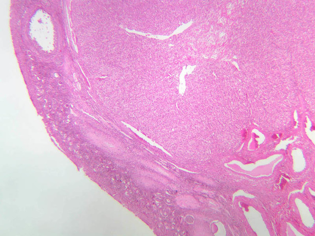
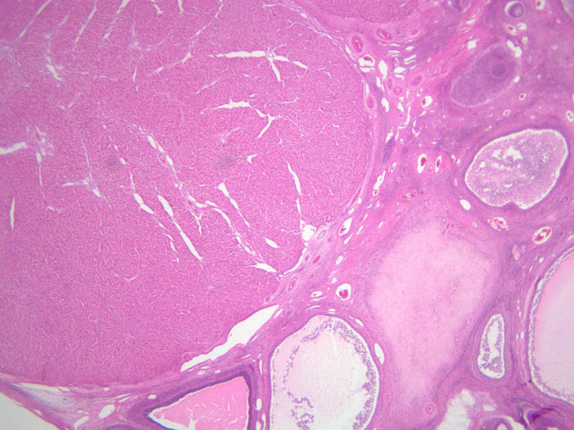
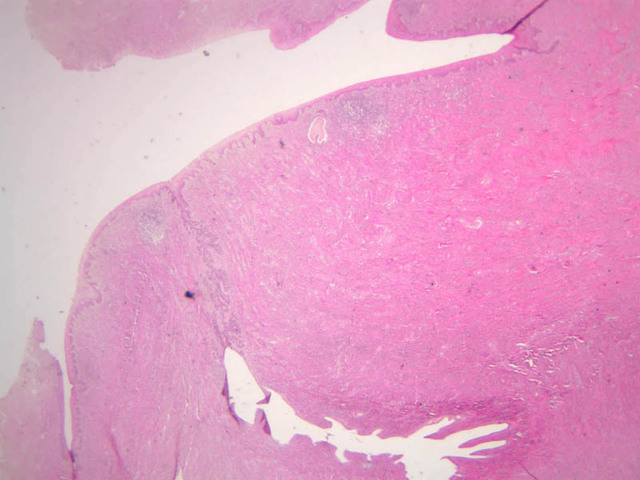
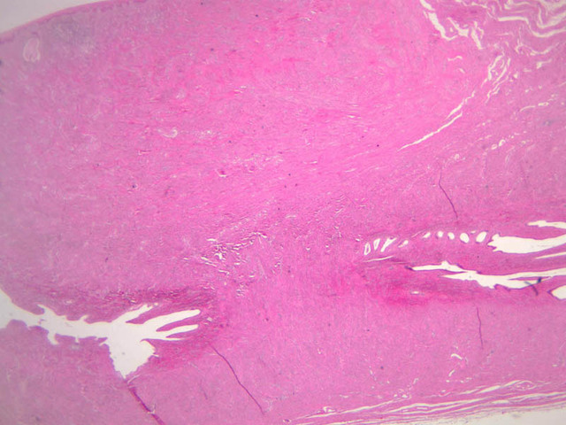
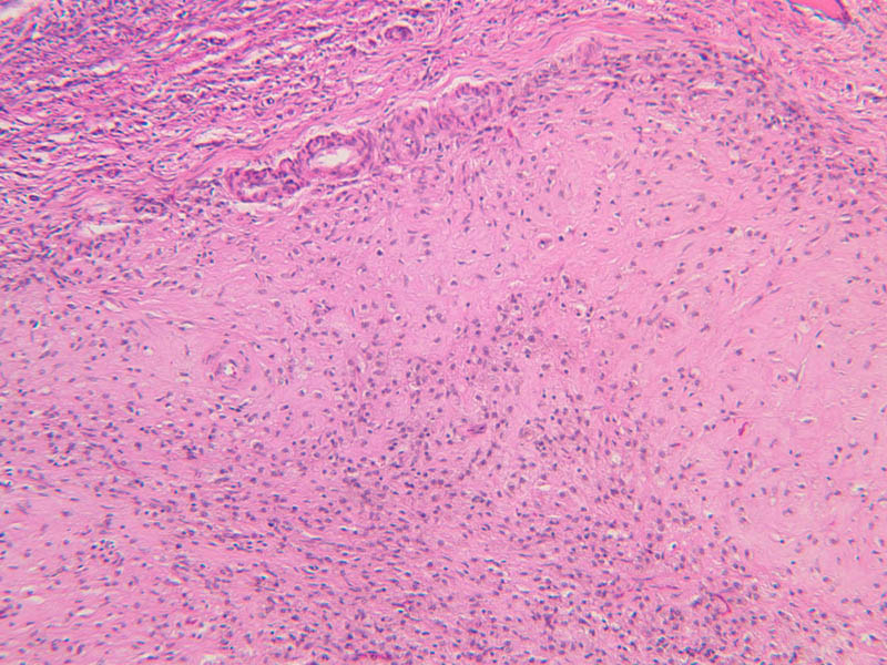
Corpus Albicans
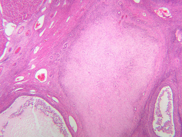
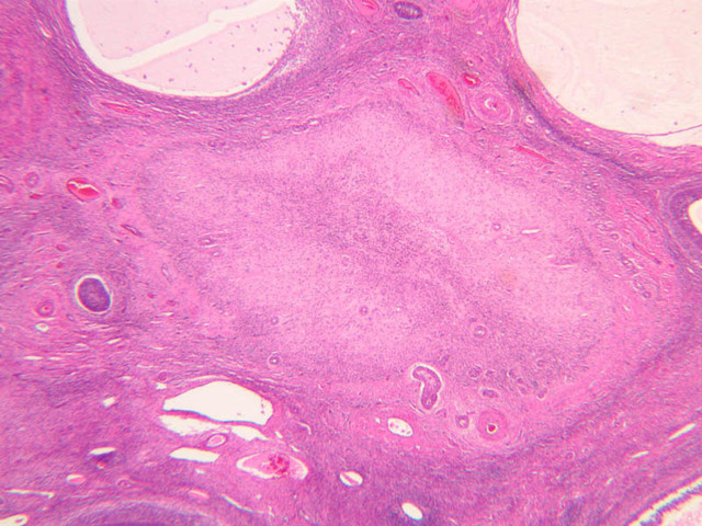
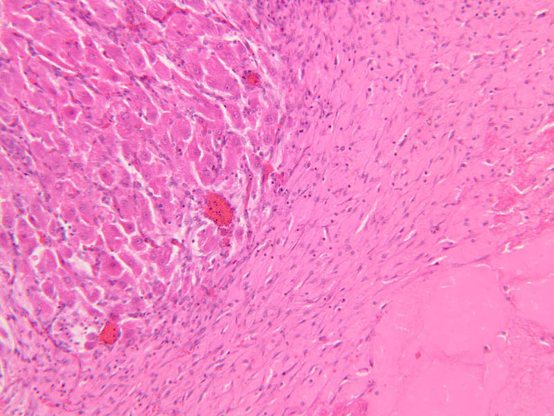
The corpus luteum persists for 12 to 14 days. If fertilized ovum does not implant, the corpus luteum degenerates, leaving a scar which is referred to as the corpus albicans. This will also disappear eventually. Some of the B-54 slides in our collection have corpus albicans – try to identify a corpus albicans on your slide or consult with your neighbors. Corpus Albicans (some B-54 [1x-labeled] [2.5x] [1x-labeled, 2.5x, 10x])Corpus Albicans Image Gallery

Corpus Albicans Table of Identifications
| Row | Structure | Abbreviation | Optimal Stain | Representative Section | Note |
|---|---|---|---|---|---|
| 1 | Corpus Luteum | (none) | H&E | |
|
| 2 | Corpus Albicans | (none) | H&E | |
|
| 3 | Medulla (OVarian) | (none) | H&E | |
|
| 4 | Cortex (Ovarian) | (none) | H&E | |
Top of page
Atretic Follicle
At any point in their development, follicles may cease maturation and undergo a process of degeneration called atresia. The process of atresia occurs through apoptotic cell death. The appearance of such follicles will depend on the stage at which atresia began as well as the extent to which it has occurred. Atretic Follicles (slides B-94 [2.5x, 10x, 20x] [10x, 10x]; B-95 [10x, 20x])Atretic Follicle Image Gallery


Top of page
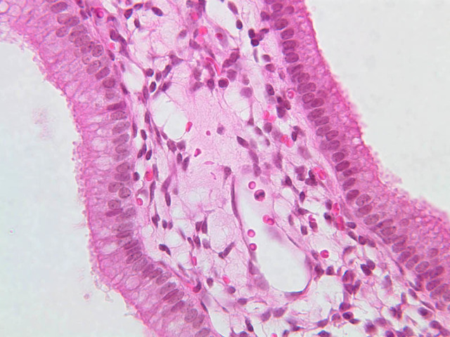
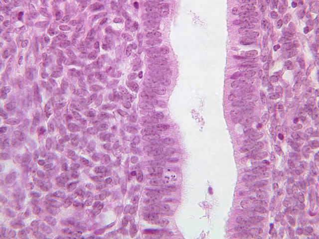
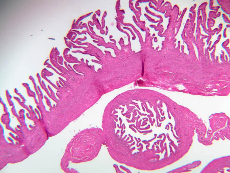
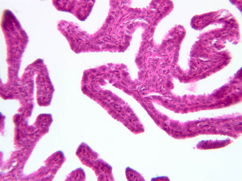
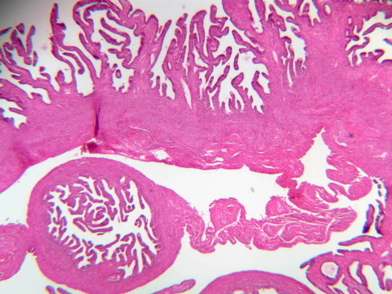
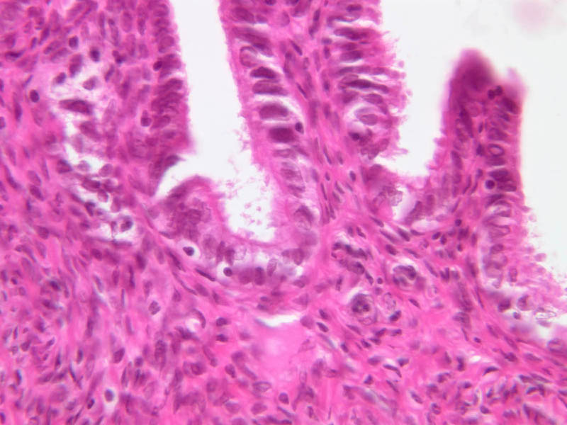
Female Duct System
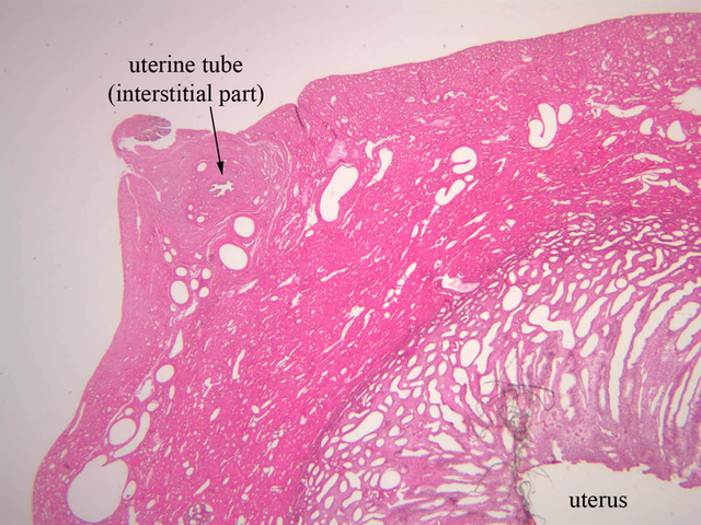
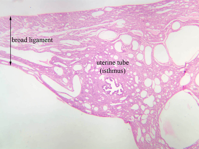
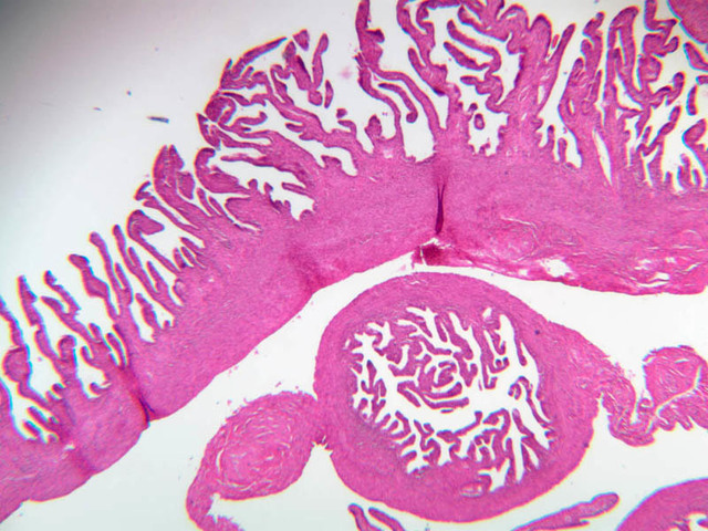
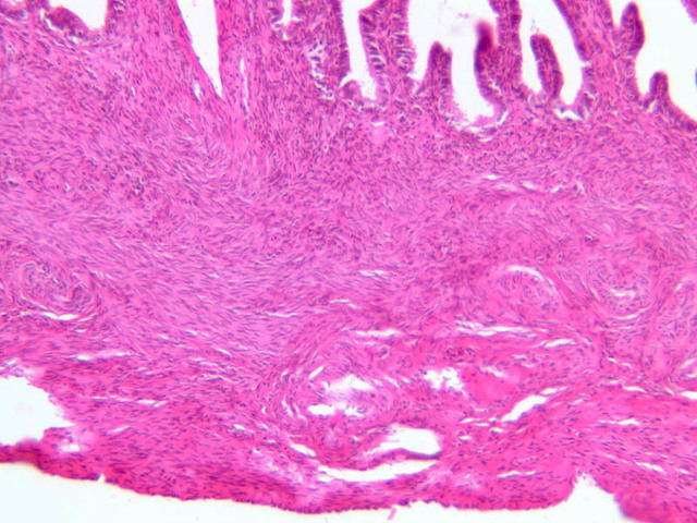
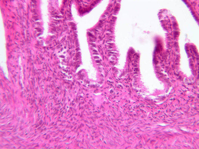
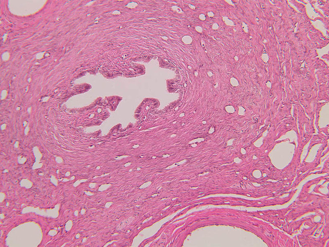
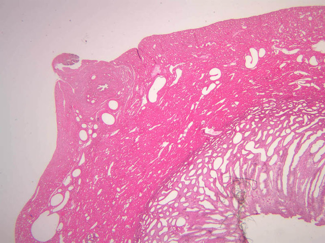
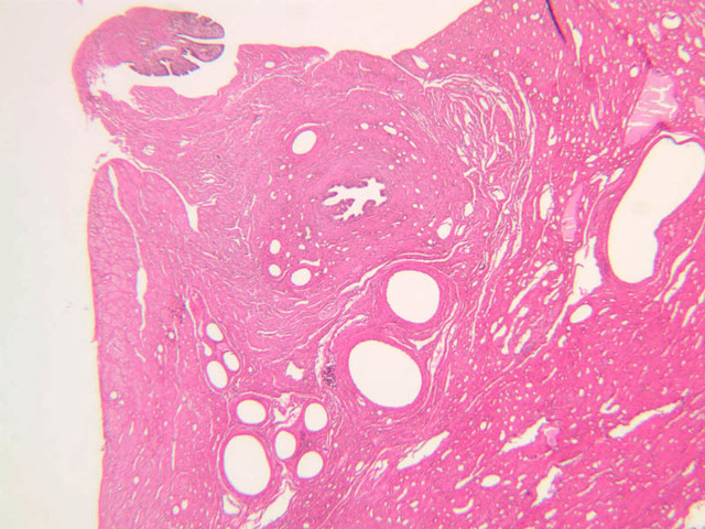
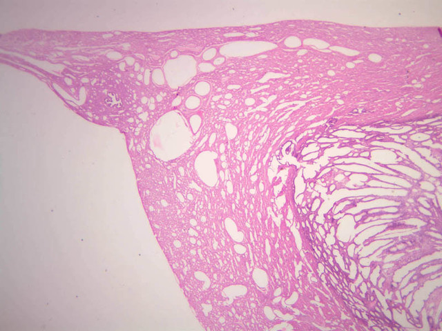
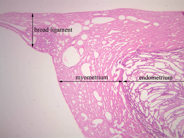
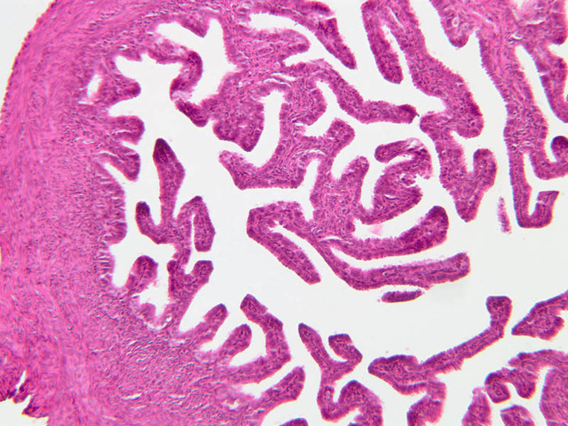
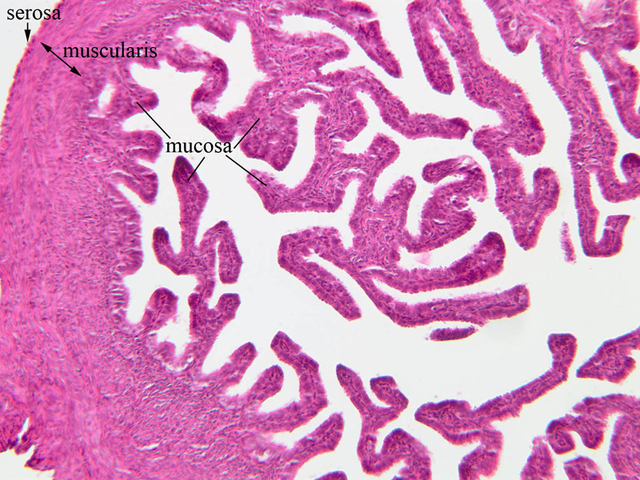
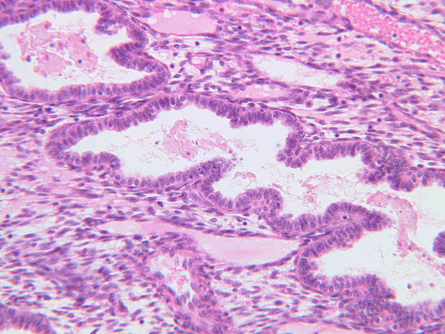
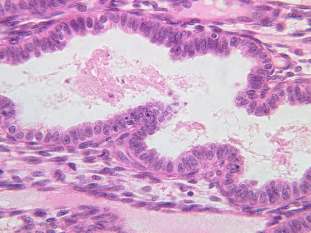
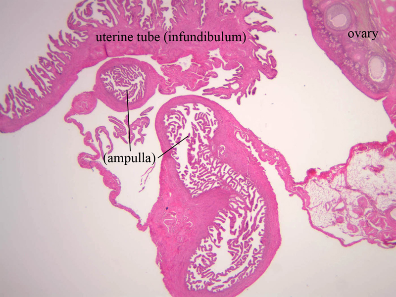
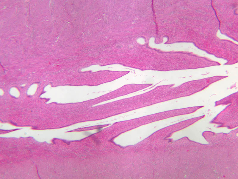
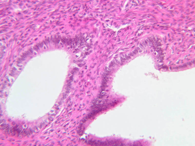
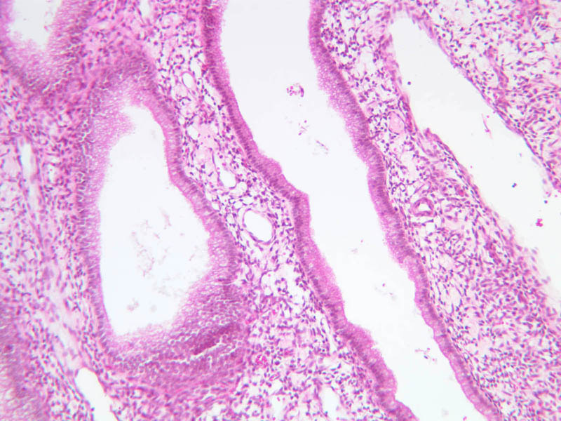
After ovulation, the oocyte passes into the uterine tube (i.e. oviduct, i.e. fallopian tube). Fertilization takes place here. This tube is subdivided into four regions:- uterine/intramural (that portion within the uterine wall) (slide B-93 [1x-labeled, 2.5x, 10x] [1x, 1x, 1x, 1x])
- isthmus (short narrow portion next to uterus) (B-93 [2.5x-labeled])
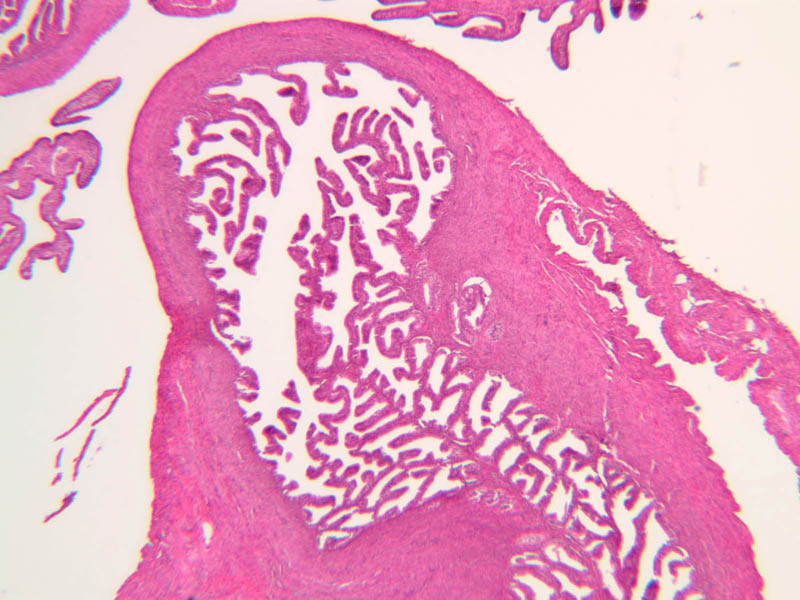
- ampulla (expanded, longest portion) (B-96 [1x-labeled, 2.5x])
- infundibulum (funnel-shaped abdominal opening) (B-96 [1x-labeled]).
Female Duct System Image Gallery


Female Duct System Table of Identifications
| Row | Structure | Abbreviation | Optimal Stain | Representative Section | Note |
|---|---|---|---|---|---|
| 1 | Uterine Tube (Interstitial) | (none) | H&E | |
|
| 2 | Uterus | (none) | H&E | |
|
| 3 | Broad Ligament | (none) | H&E | |
|
| 4 | Uterine Tube (Isthmus) | (none) | H&E | |
|
| 5 | Uterine Tube (Infundibulum) | (none) | H&E | |
|
| 6 | Uterine Tube (Ampulla) | (none) | H&E | |
|
| 7 | Ovary | (none) | H&E | |
|
| 8 | Serosa | (none) | H&E | |
|
| 9 | Muscularis | (none) | H&E | |
|
| 10 | Mucosa | (none) | H&E | |
|
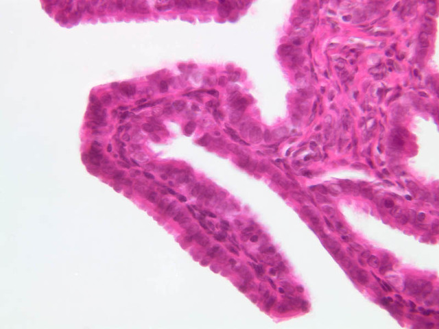
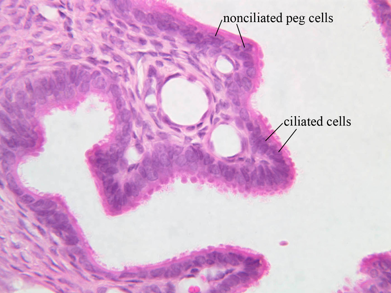
| 11 | Nonciliated Peg Cells | (none) | H&E | |
|
| 12 | Ciliated Cells | (none) | H&E | |
Top of page
Uterus
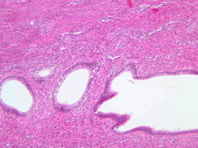
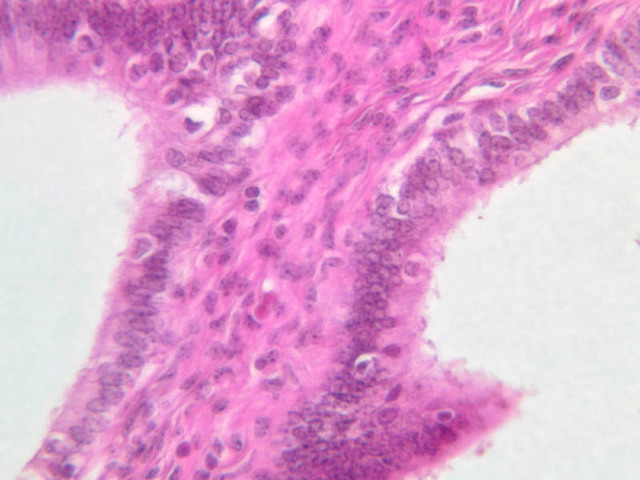
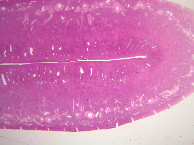
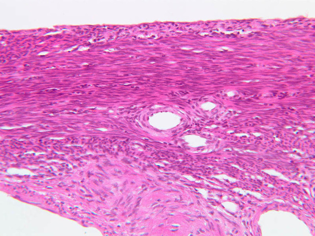
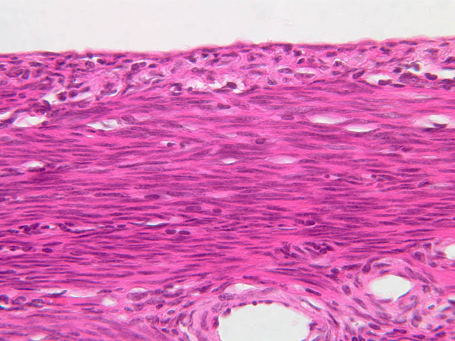
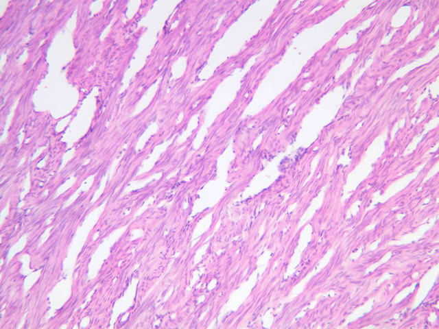
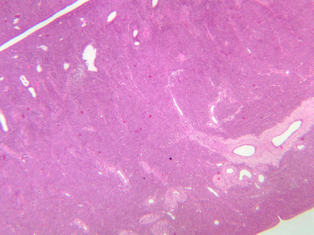
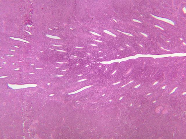
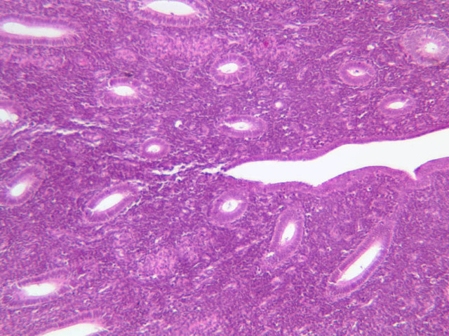
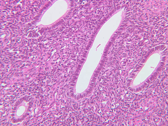
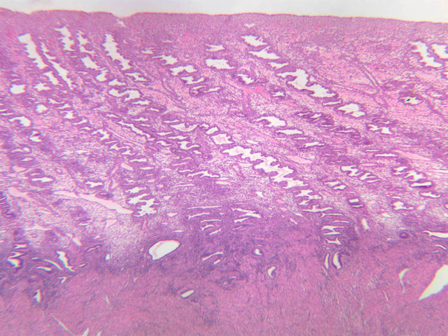
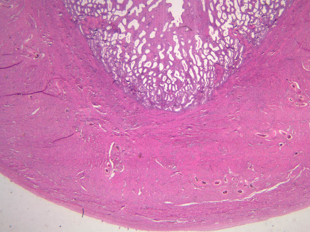
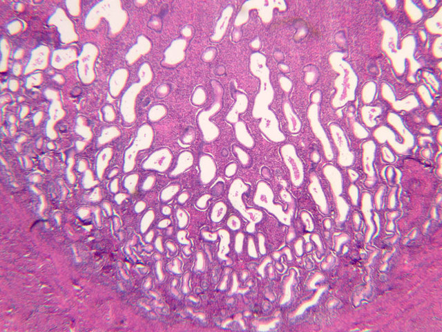
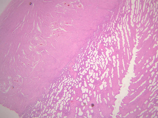
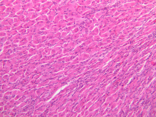
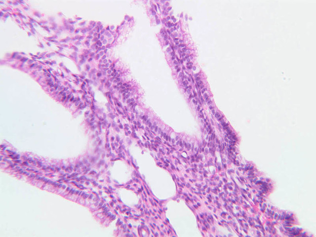
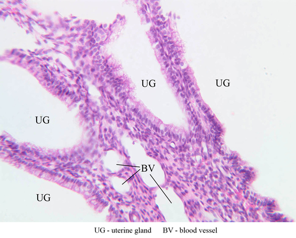
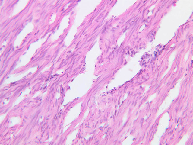
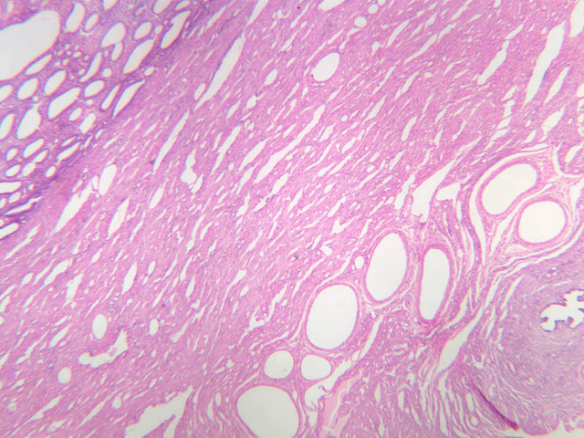
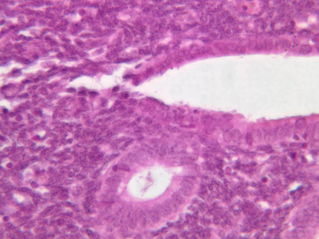
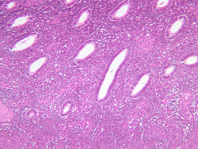
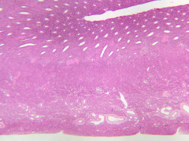
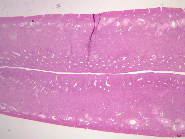
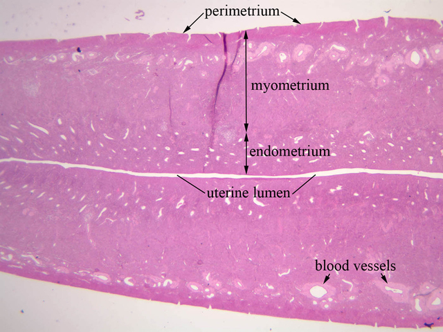
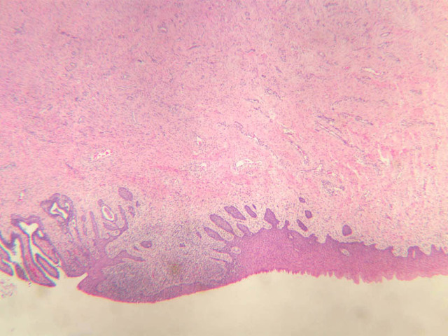
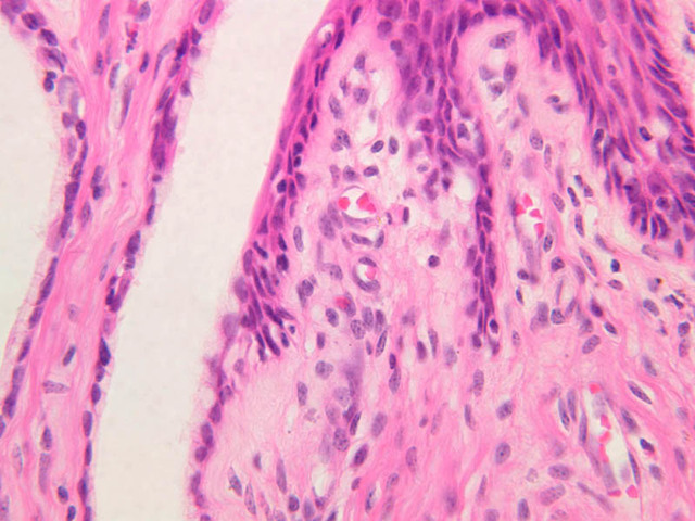
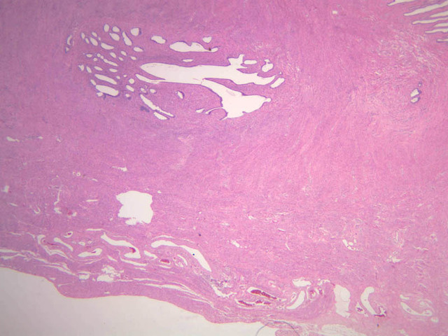
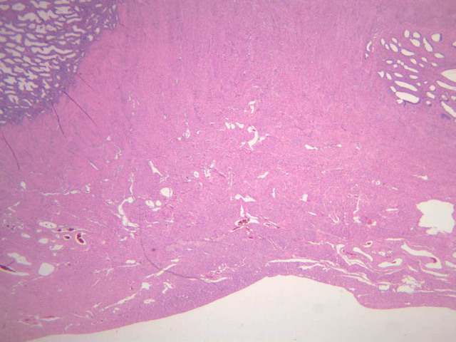
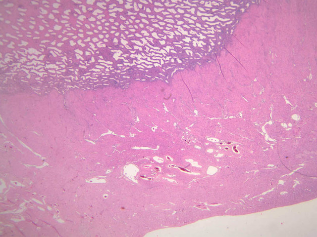
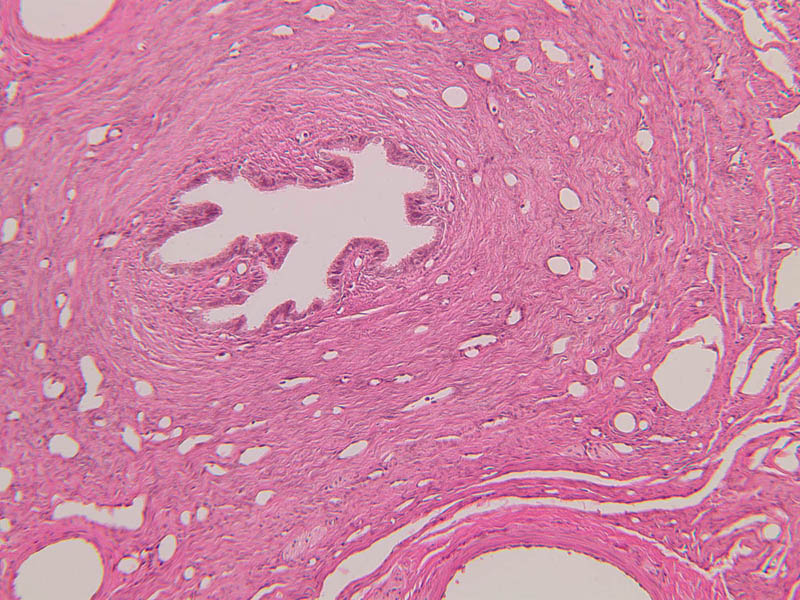
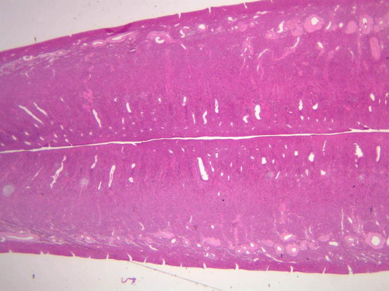
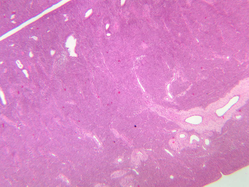
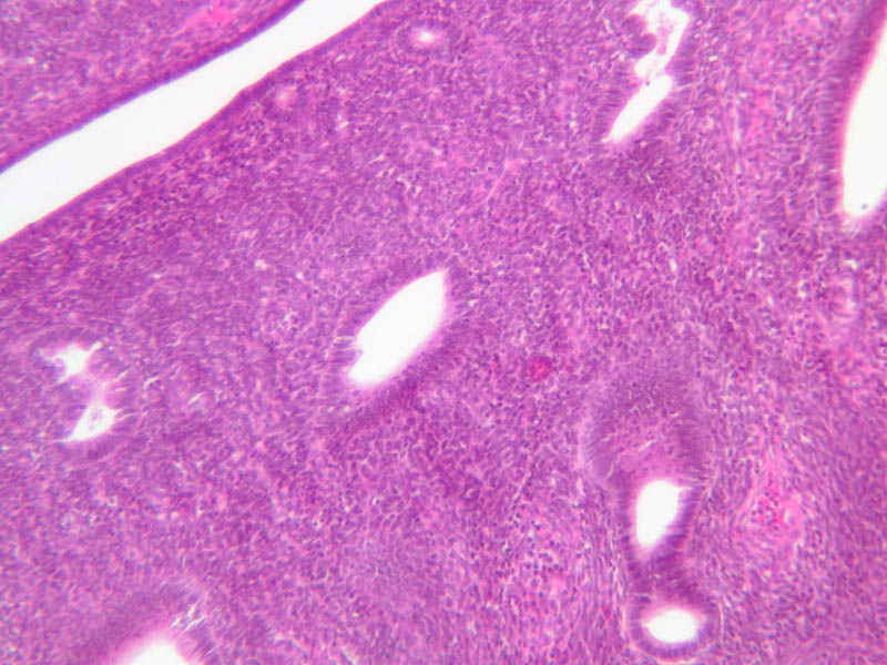
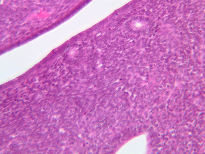
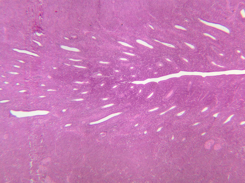
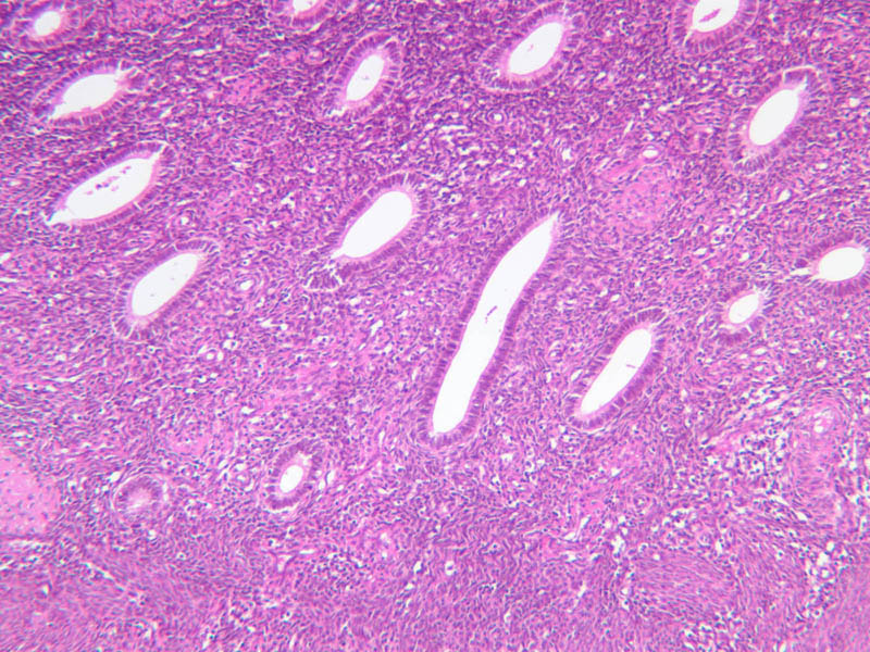
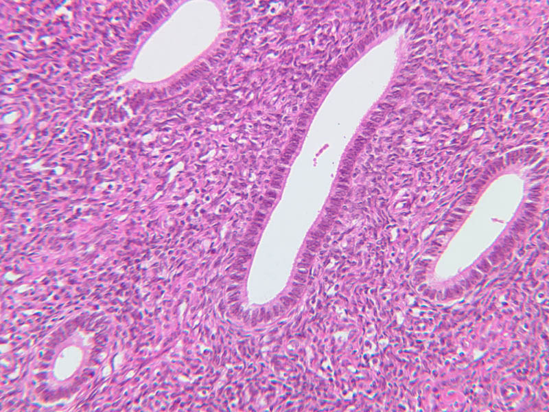
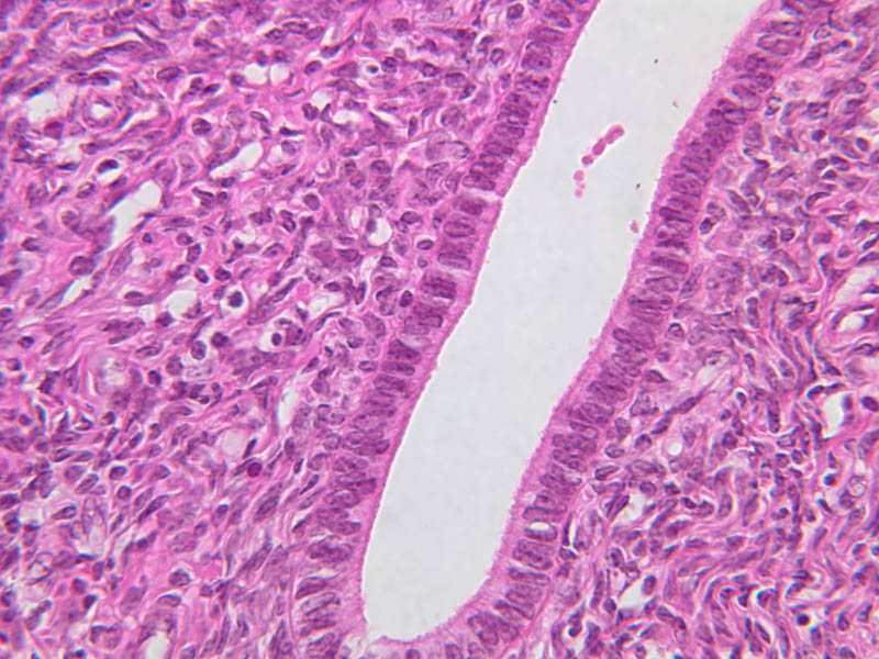
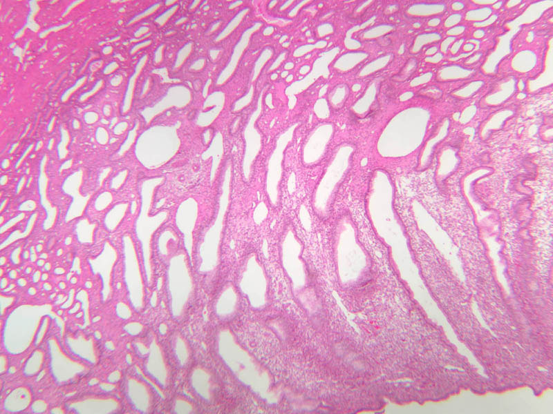
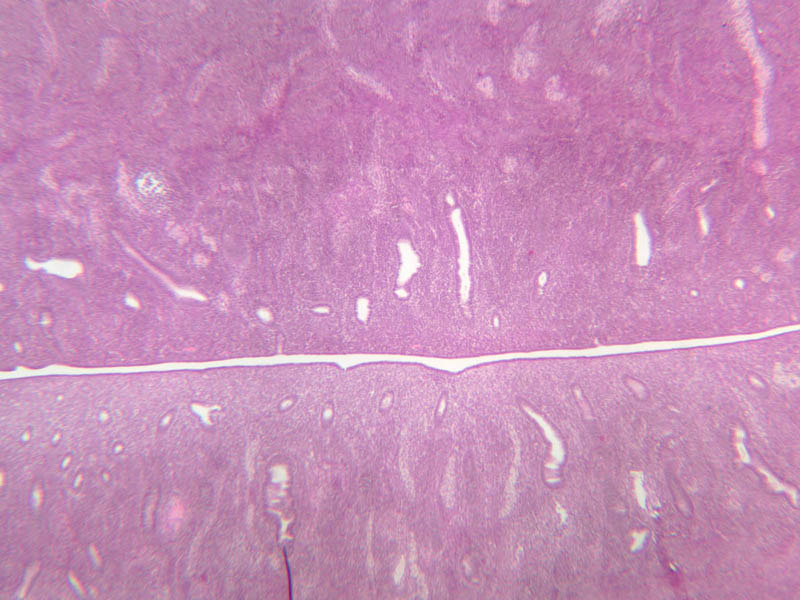
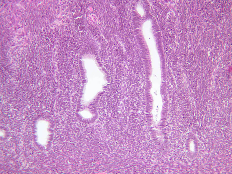
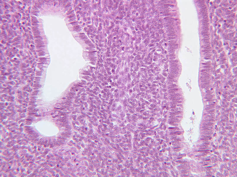
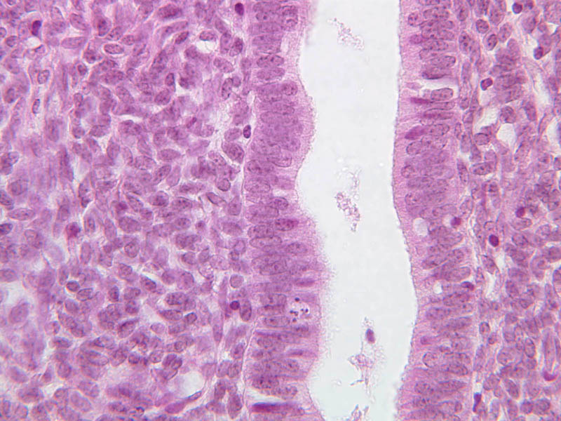
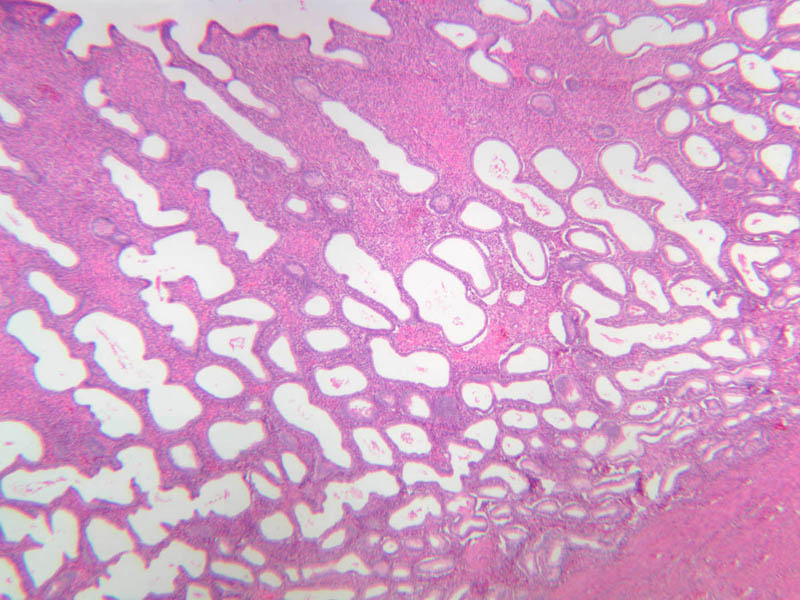
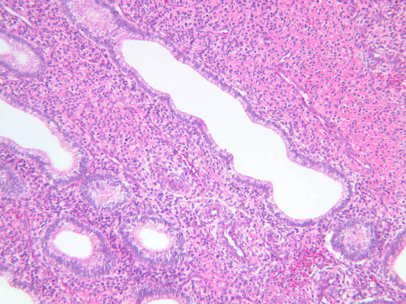
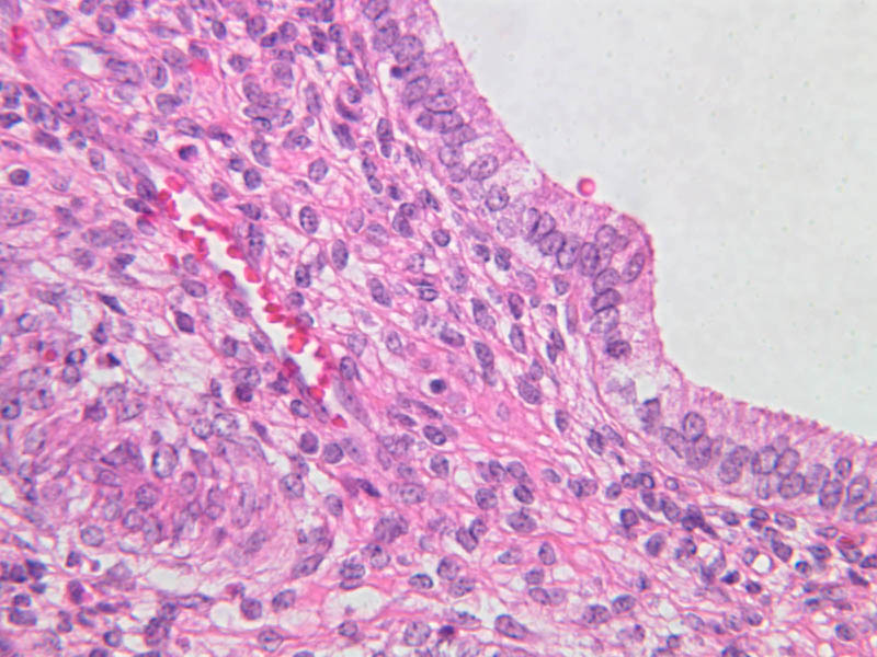
The uterus is a pear-shaped muscular organ, somewhat flattened from front to back. The expanded upper portion is referred to as the fundus, while its lower part, which protrudes into the vagina, is called the cervix. The lumen of the uterus is slit-like and is in communication with that of the uterine tubes (B-97, monkey uterus [1x, 1x, 1x, 1x]). The wall of the uterus consists of three layers: perimetrium, myometrium, and endometrium (B-97 [1x, 1x-labeled] [1x, 1x, 1x]; B-100 [1x, 1x]). The perimetrium is peritoneum which covers the uterus, but is lacking in the posterocaudal third below the peritoneal reflection (B-97 [2.5x, 10x, 20x, 40x]). The myometrium is the very thick middle layer consisting of bundles of smooth muscle cells arranged into several interwoven layers (slide B-93 [1x-labeled, 2.5x] [1x, 2.5x] [1x, 2.5x] [2.5x, 10x, 20x, 40x]). The endometrium is the innermost layer and is a glandular mucosa (B-97 [2.5x, 10x, 20x] [2.5x, 10x, 20x] [2.5x, 10x, 20x, 40x]). It consists of a surface epithelium which is invaginated into tubes called uterine glands. The glands penetrate into a very thick lamina propria referred to as the endometrial stroma. The surface epithelium is a mixture of ciliated and non-ciliated simple columnar cells, whereas the glandular epithelium consists mainly of non-ciliated secretory cells. The endometrial stroma has the appearance of a loose, rather cellular mesenchyme with numerous blood vessels. The endometrium is subdivided into two main parts; the basalis and the functionalis (slide B-97). The basalis is deep, lying adjacent to the myometrium, it consists of stroma and the closed ends of the uterine glands and is not sloughed during menstruation. The functionalis is upper zone consisting of the remainder of the uterine glands with intervening stroma. It is lost during menstruation, but is replaced again with each menstrual cycle, by proliferation of the persisting elements in the basalis. The menstrual cycle is a continuous series of events, dominated by the endocrine activity of the ovary, which repeats itself with a 28 day period. The endometrium participates by undergoing a series of structural changes which are roughly divided into three categories, the proliferative, secretory, and menstrual phases. These phases are listed below, examine each of the uterine slides and determine to which phase it belongs.Uterus Image Gallery


Uterus Table of Identifications
| Row | Structure | Abbreviation | Optimal Stain | Representative Section | Note |
|---|---|---|---|---|---|
| 1 | Perimetrium | (none) | H&E | |
|
| 2 | Myometrium | (none) | H&E | |
|
| 3 | Endometrium | (none) | H&E | |
|
| 4 | Uterine Lumen | (none) | H&E | |
|
| 5 | Blood Vessels | (none) | H&E | |
|
| 6 | Broad Ligament | (none) | H&E | |
Top of page
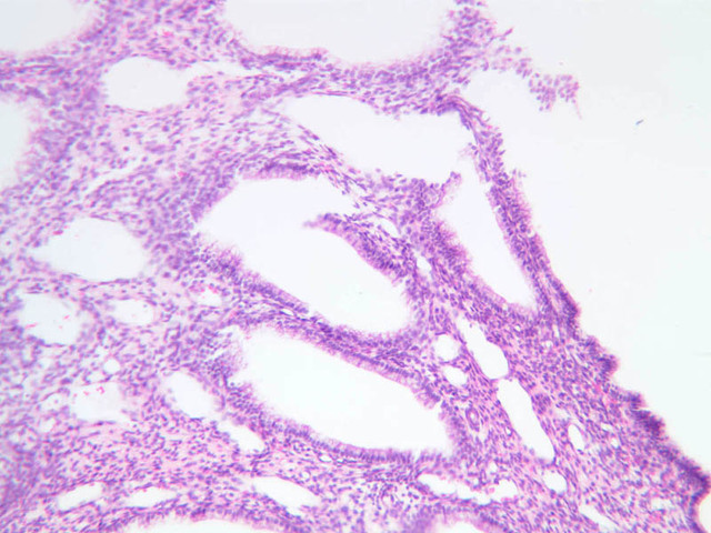
Menstrual Phase
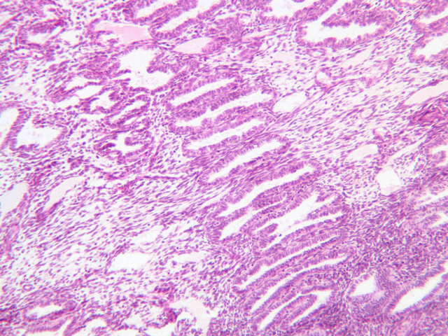
The menstrual phase lasts 1 -5 days, and corresponds to ovarian menstrual phase; i.e., degeneration of corpus luteum with consequent decrease in estrogen and progesterone levels. During this phase the functionalis sloughs off leaving the basalis. Just prior to sloughing, the glands are extremely dilated and tortuous.Proliferative Phase
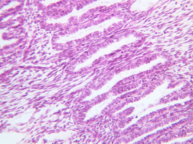
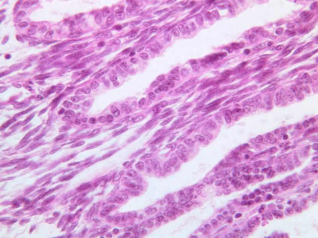
This phase occurs during days 7 to 14 and corresponds to the ovarian follicular phase with rising levels of estrogens. The functionalis layer is replaced by growth of the endometrial elements in the basalis. The glands are usually straight tubes extending from the surface. Epithelial cells become taller and accumulate glycogen basal to their nuclei. (slide B-93 [2.5x, 10x, 20x, 40x]; B-97 [2.5x, 10x, 20x, 40x])Proliferative Phase Image Gallery


Top of page
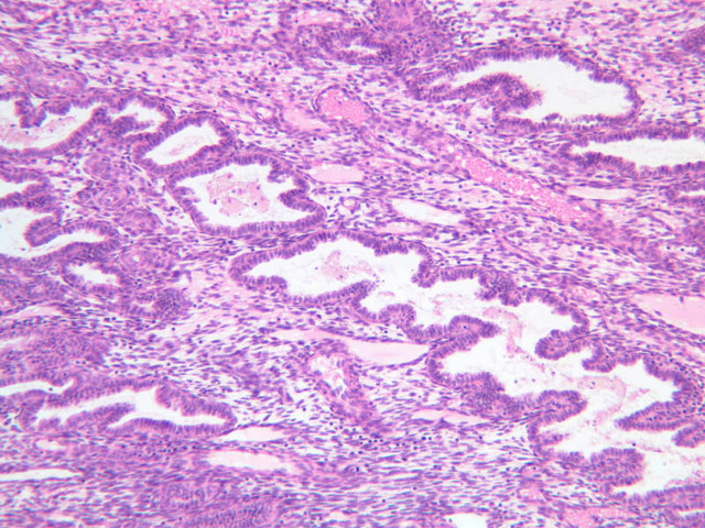
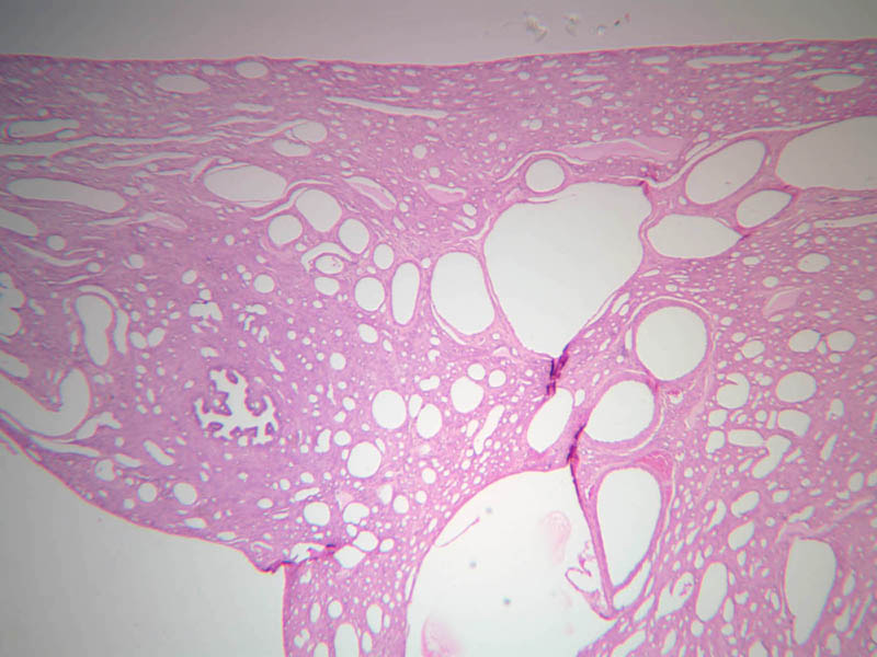
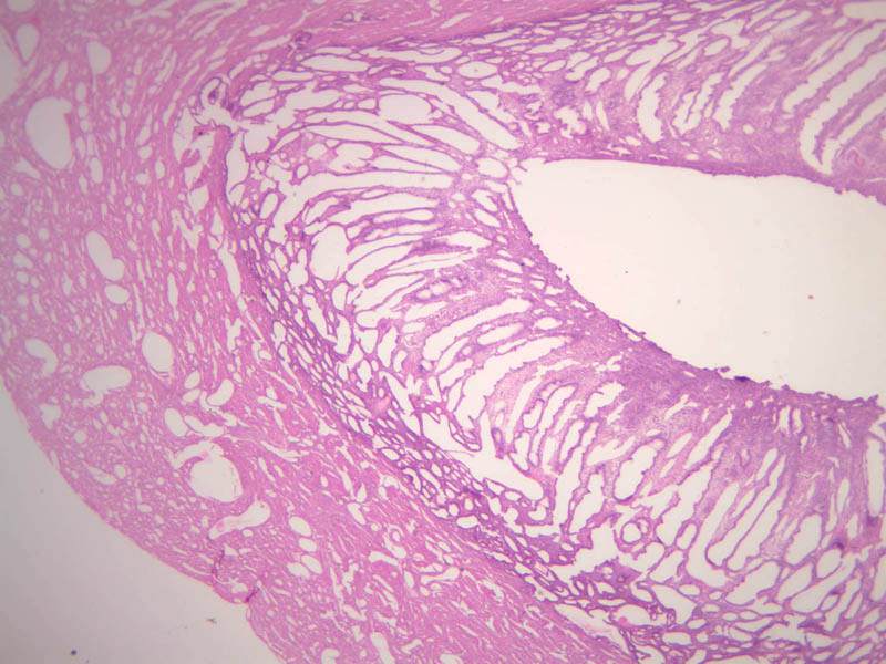
Secretory Phase
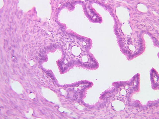
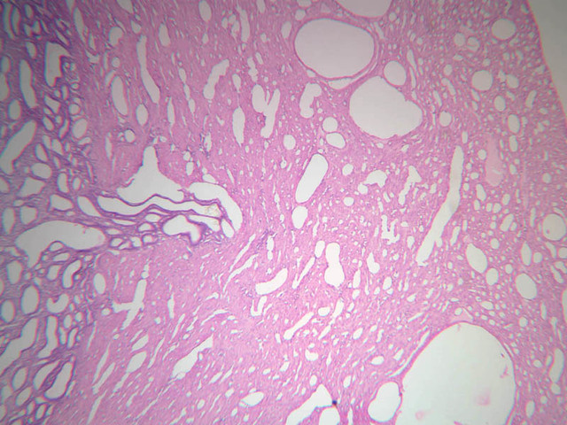
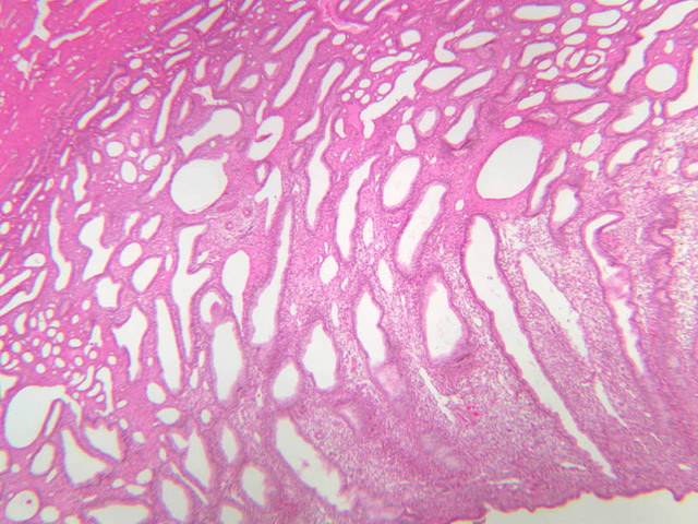
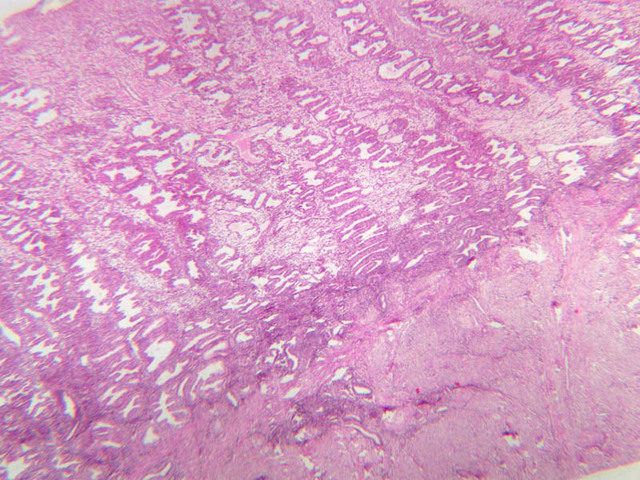
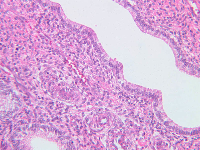
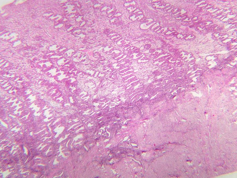
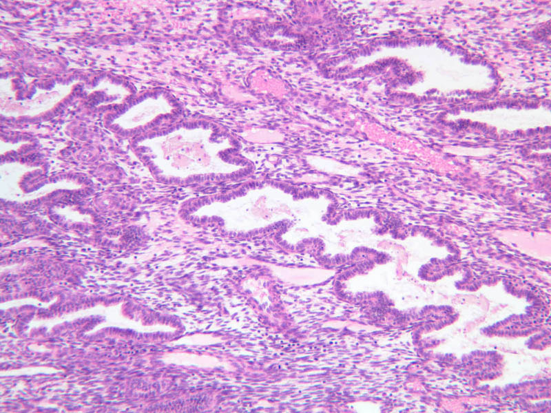
This phase occurs on days 15 to 27 and corresponds to the ovarian luteal phase characterized by rising levels of progesterone. The epithelial cells begin to secrete a mucoid fluid rich in nutrients, especially glycogen. The glands become highly coiled and folded and toward the end, very distended. The density of the stroma lessens as it becomes edematous. (slides B-99 [2.5x, 10x, 20x, 40x] [2.5x, 10x, 20x, 40x]; B-100 [1x, 2.5x] [2.5x, 10x, 20x, 40x] [2.5x, 10x, 20x, 40x])Secretory Phase Image Gallery


Top of page
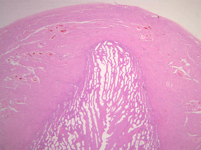
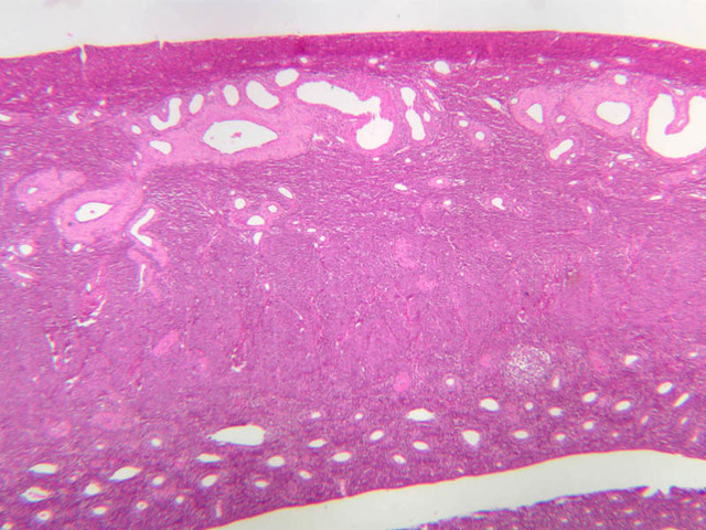
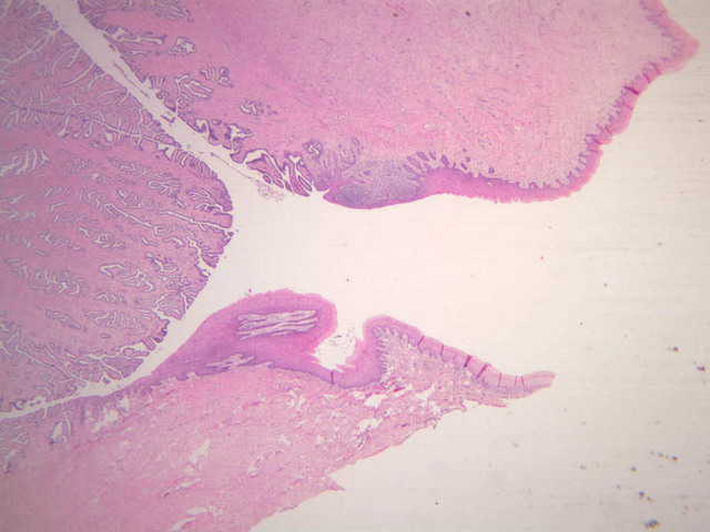
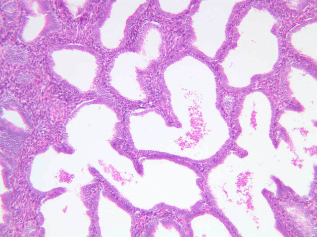
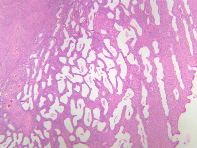
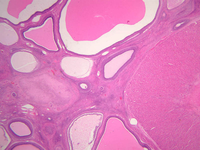
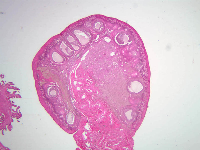
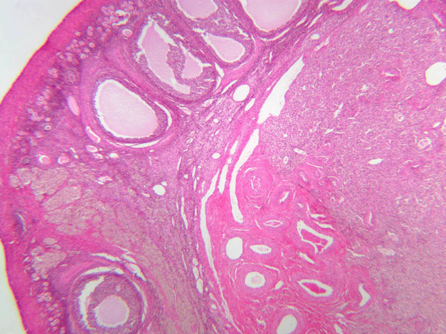
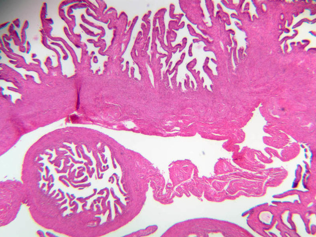
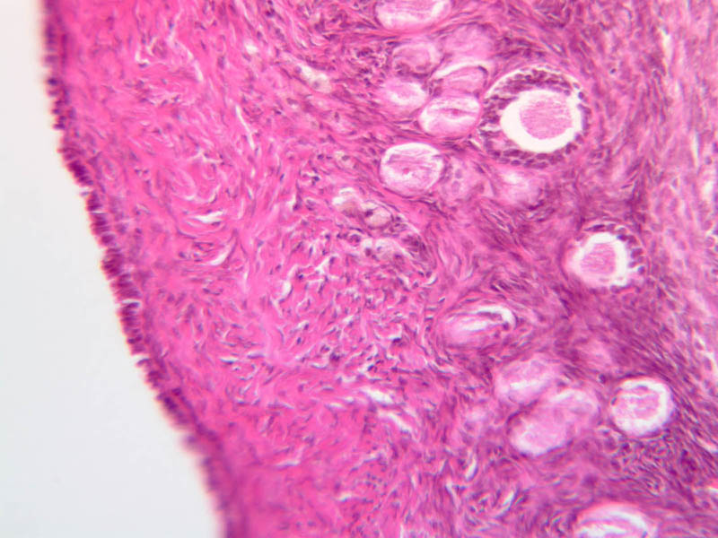
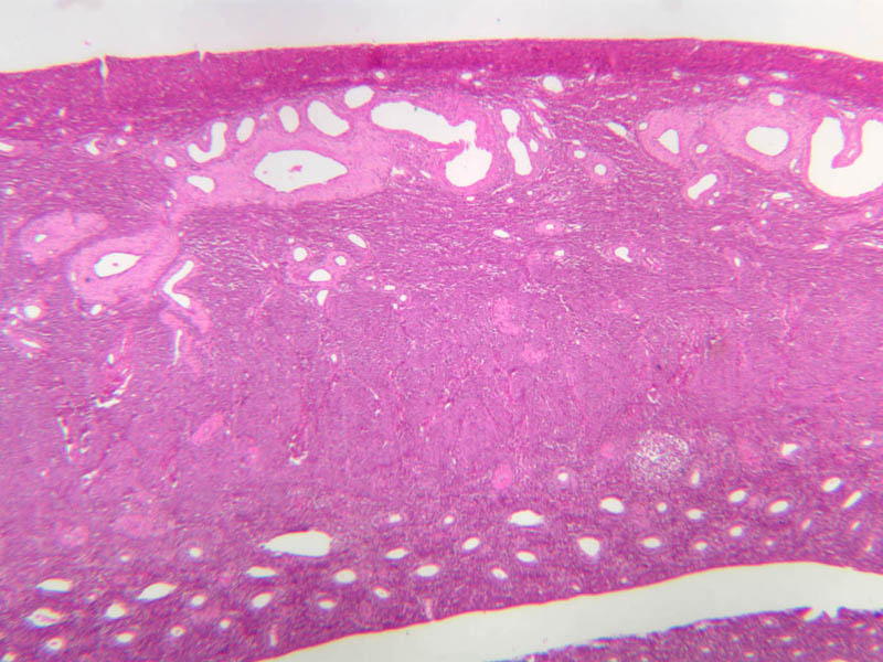
Uterine Cervix
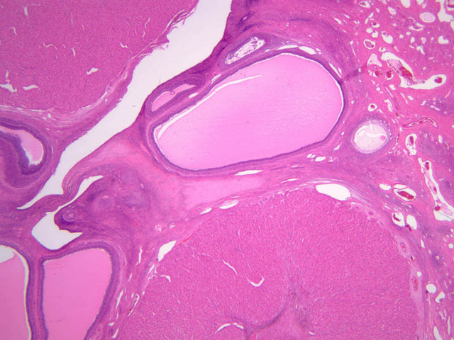
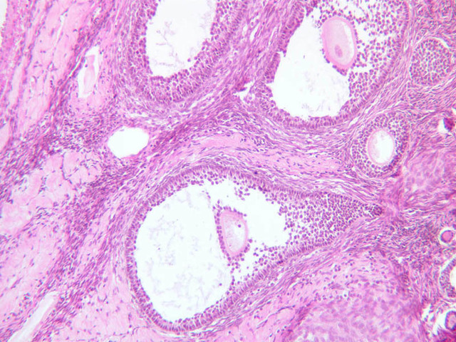
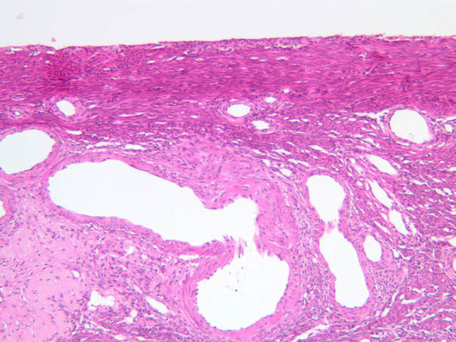
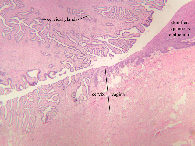
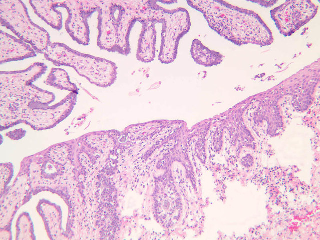
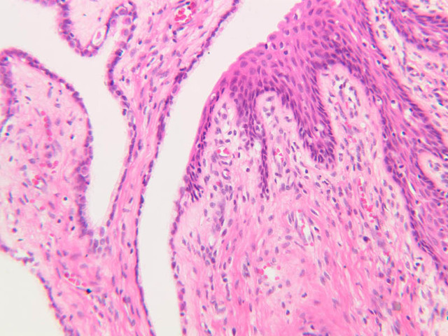
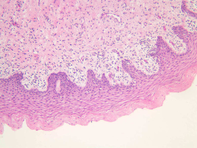
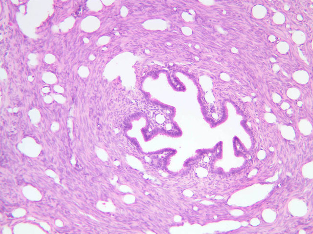
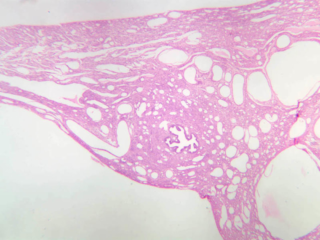
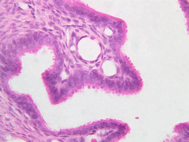
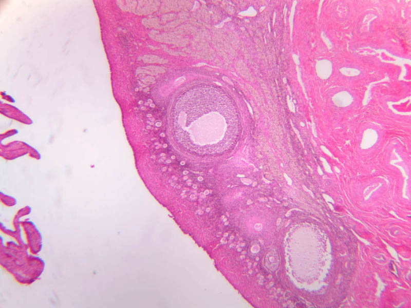
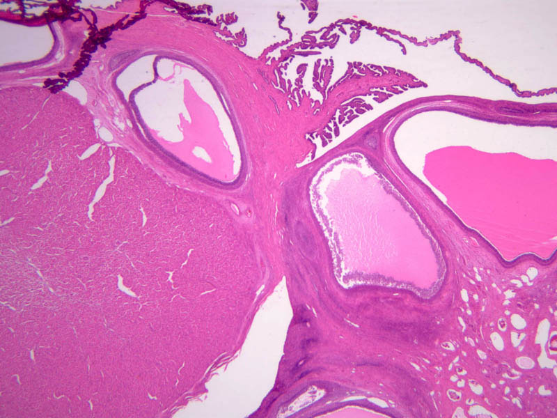
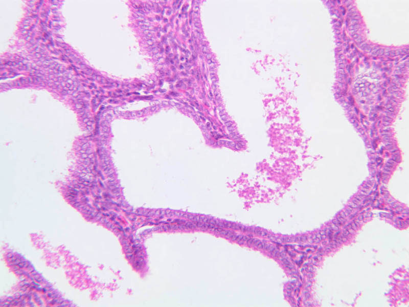
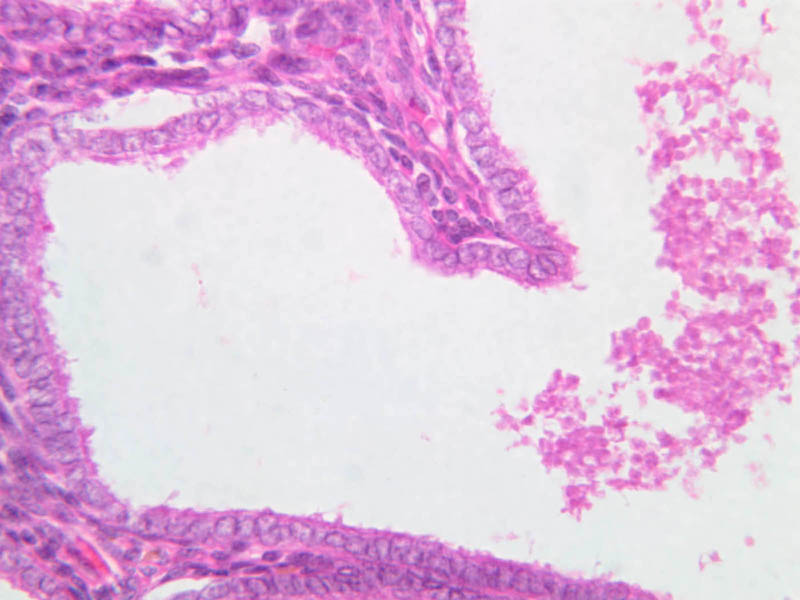
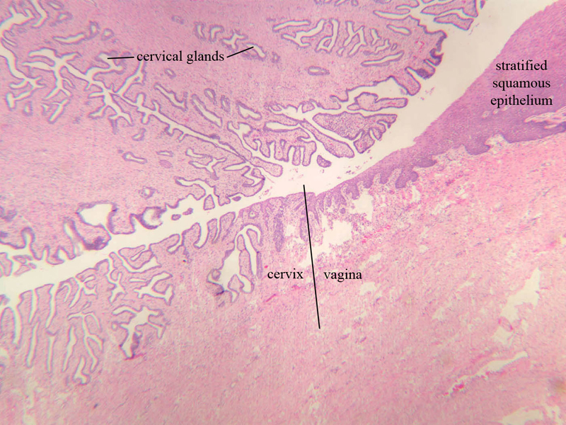
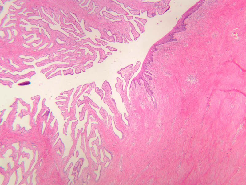
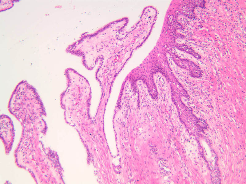
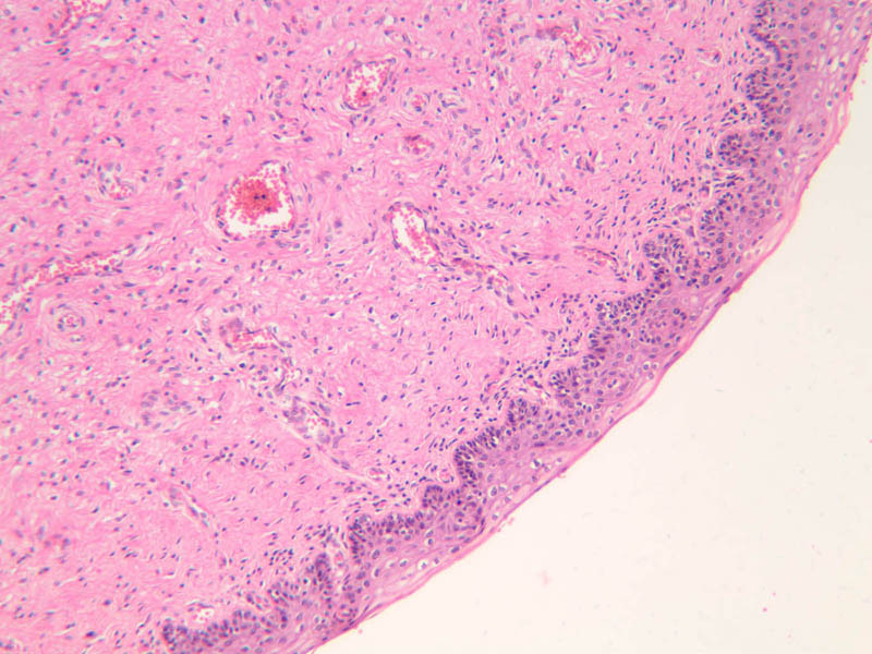
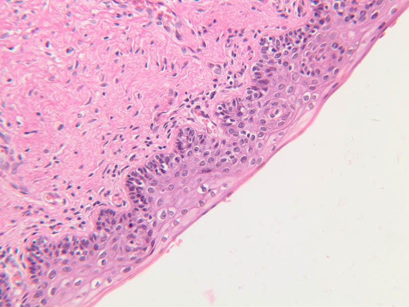
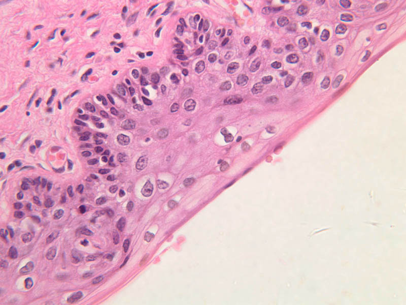
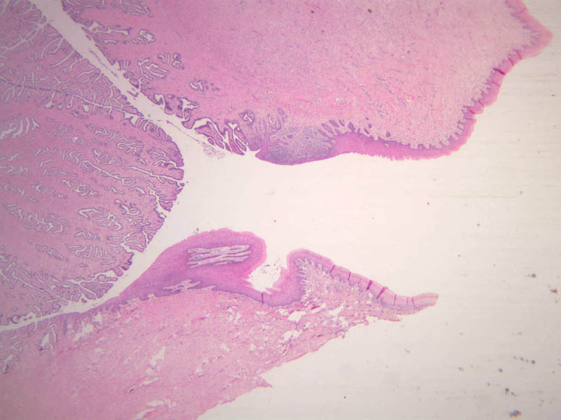
The endometrium of the cervical canal does not slough during the menstrual phase. The glands in this region are relatively large and extensively branched. They are lined by a tall simple columnar epithelium very active in the secretion of mucus. Occasionally these glands become blocked forming Nabothian cysts. The transition of the epithelium of the cervix to that of the vagina is abrupt and is in the region of the external os. At this point, the cervical simple columnar is replaced by the vaginal stratified squamous epithelium (slide B-98 [2.5x-labeled, 10x, 20x, 40x] [2.5x, 10x, 20x, 40x]). This is an area which is commonly inflamed as well as a primary location of cervical cancer.Uterine Cervix Image Gallery


Uterine Cervix Table of Identifications
| Row | Structure | Abbreviation | Optimal Stain | Representative Section | Note |
|---|---|---|---|---|---|
| 1 | Cervical Glands | (none) | H&E | |
|
| 2 | Cervix | (none) | H&E | |
|
| 3 | Vagina | (none) | H&E | |
|
| 4 | Stratified Squamous Epithelium | (none) | H&E | |
Top of page
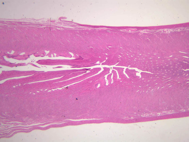
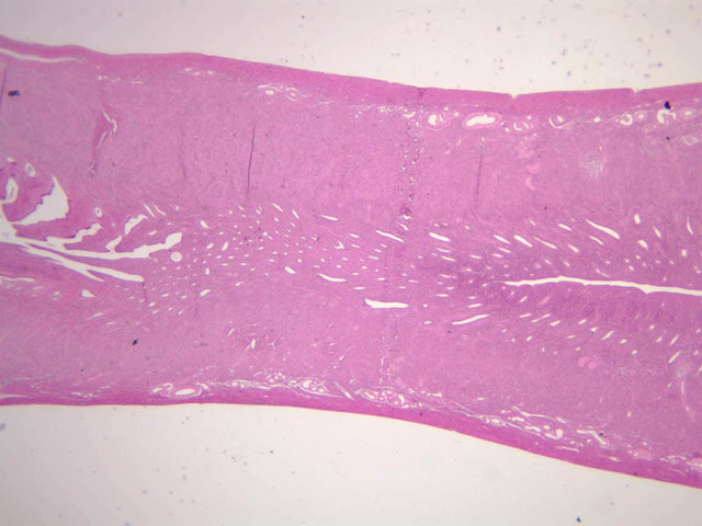
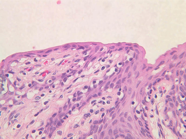
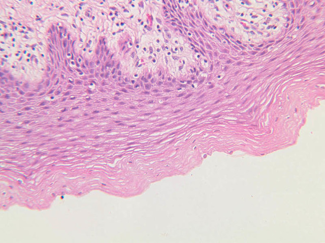
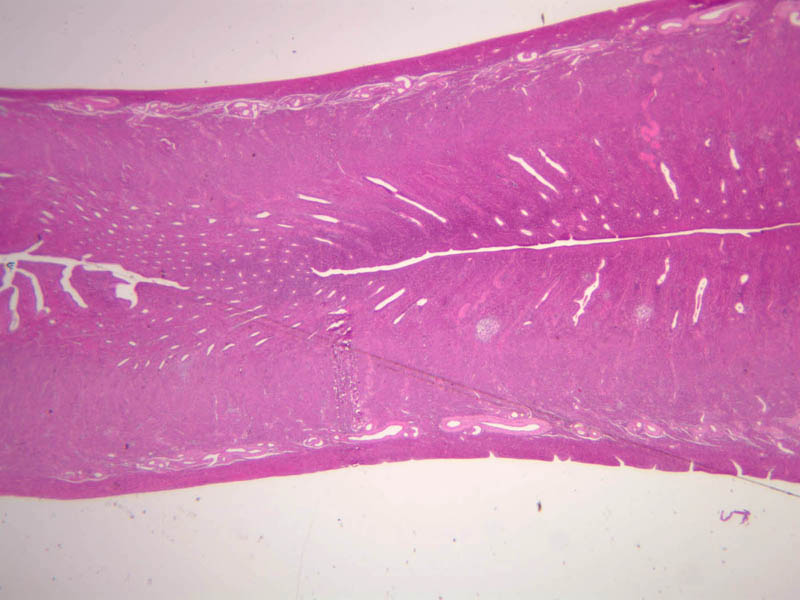
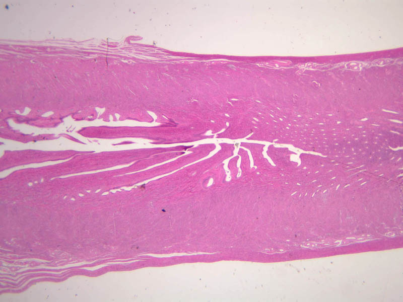
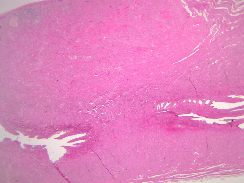
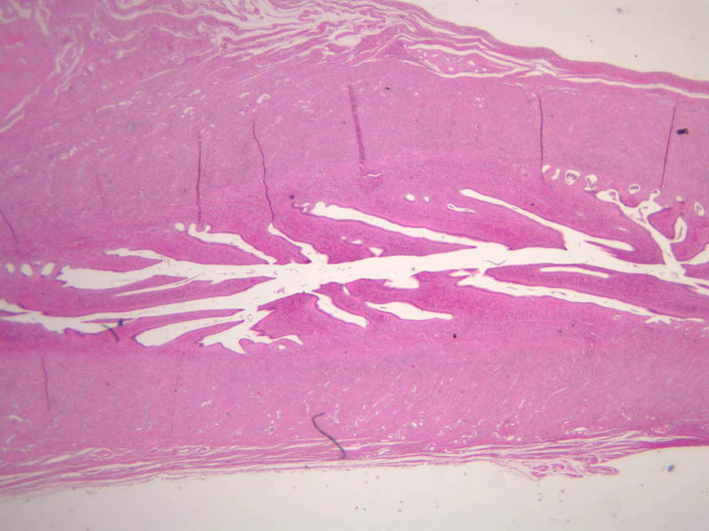
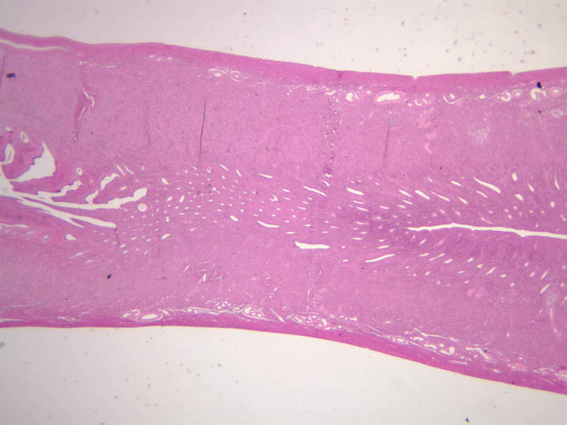
Vagina
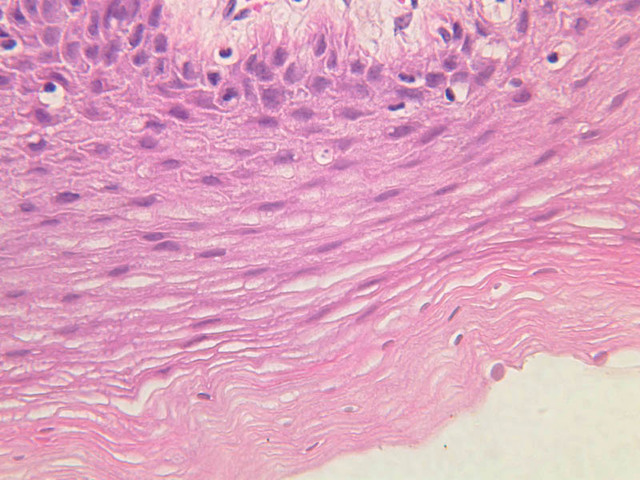
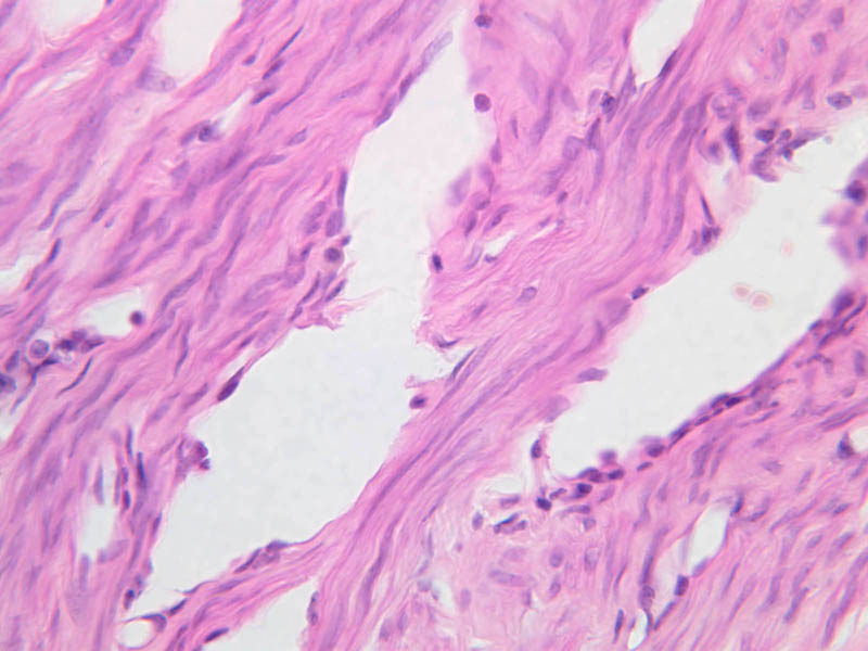
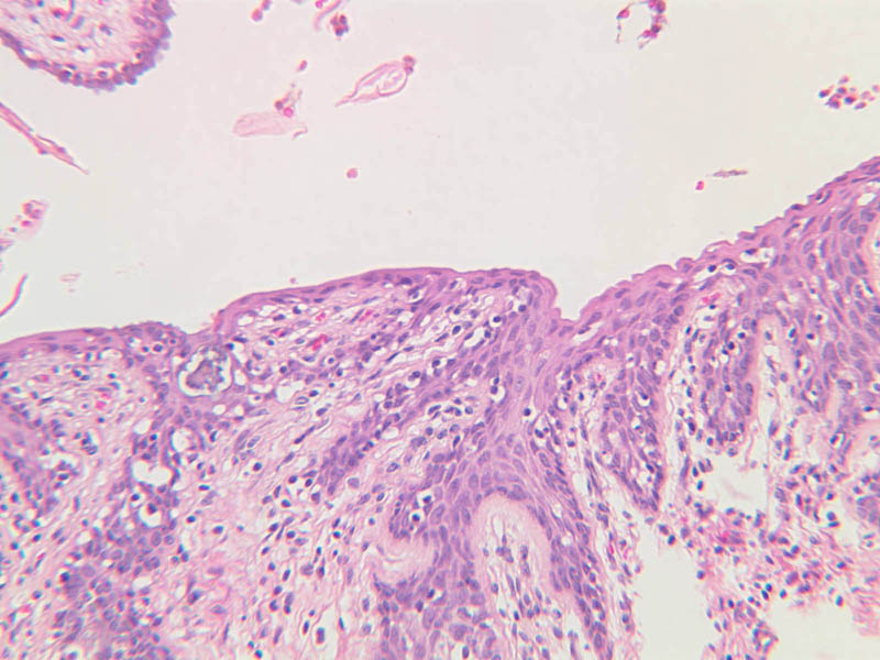
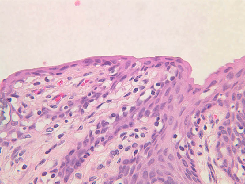
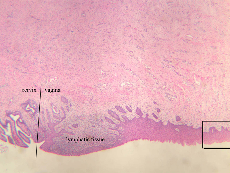
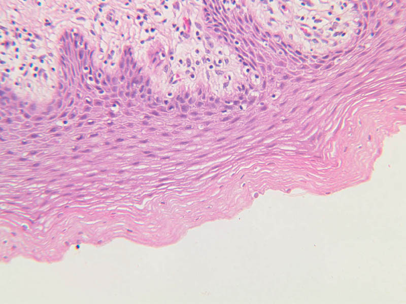
The mucosa of the vaginal wall is lined by non-keratinized stratified squamous epithelium which rests on a lamina propria. The epithelium is indented from beneath by connective tissue papillae. Occasionally aggregations of lymphocytes may be seen within the lamina propria (slide B-98 [2.5x-labeled, 10x, 20x, 40x] [2.5x, 10x, 20x, 40x] [1x, 1x, 1x, 1x, 1x]). The muscularis is said to consist of a poorly defined inner circular and outer longitudinal layer of smooth muscle. An adventitia of fibrous tissue forms the outer most layer of the vaginal wall. Although there is a rather rich vascular plexus in the lamina propria, no glands are present at any location in the vaginal wall.Vagina Image Gallery


Vagina Table of Identifications
| Row | Structure | Abbreviation | Optimal Stain | Representative Section | Note |
|---|---|---|---|---|---|
| 1 | Cervix | (none) | H&E | |
|
| 2 | Vagina | (none) | H&E | |
|
| 3 | Lymphatic Tissue | (none) | H&E | |
Top of page
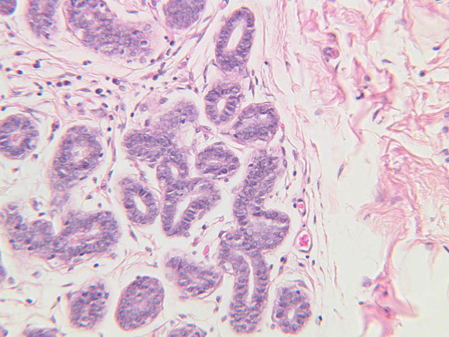
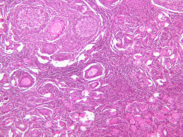
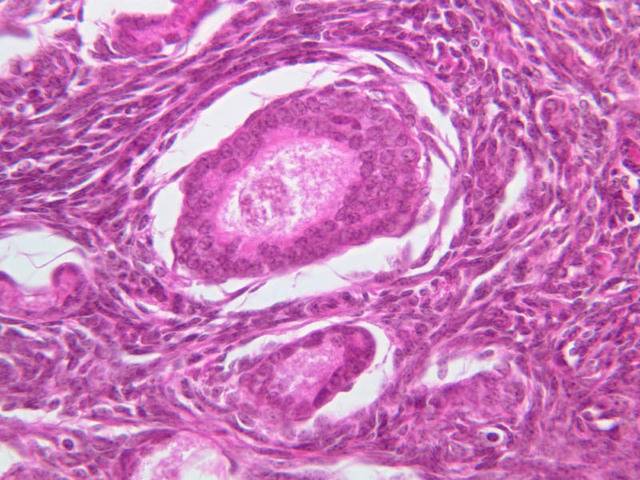
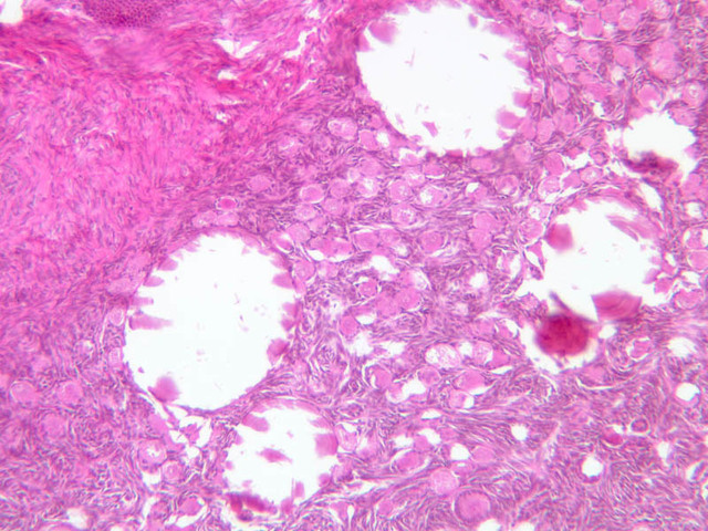
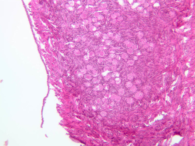
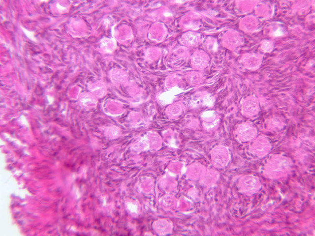
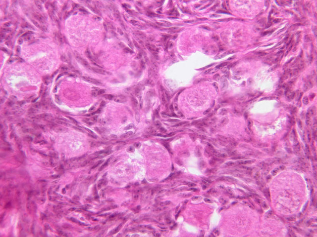
Mammary Gland
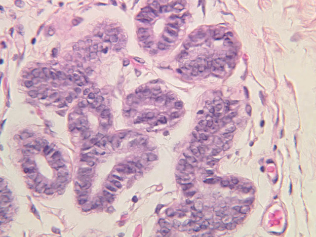
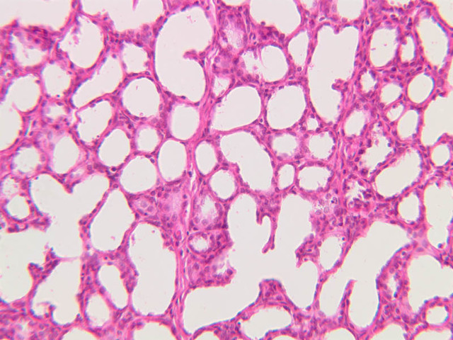
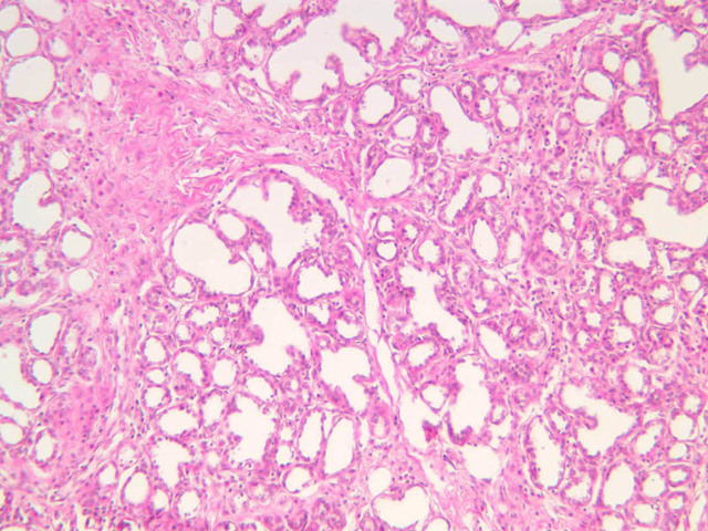
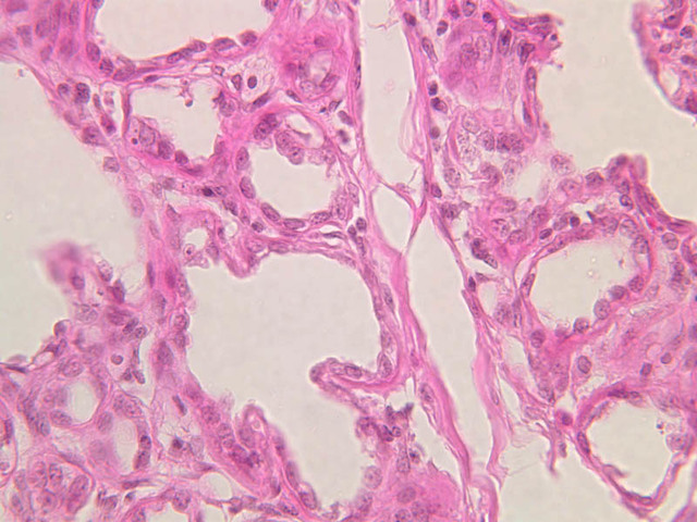
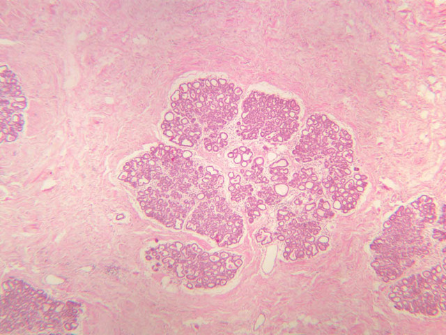
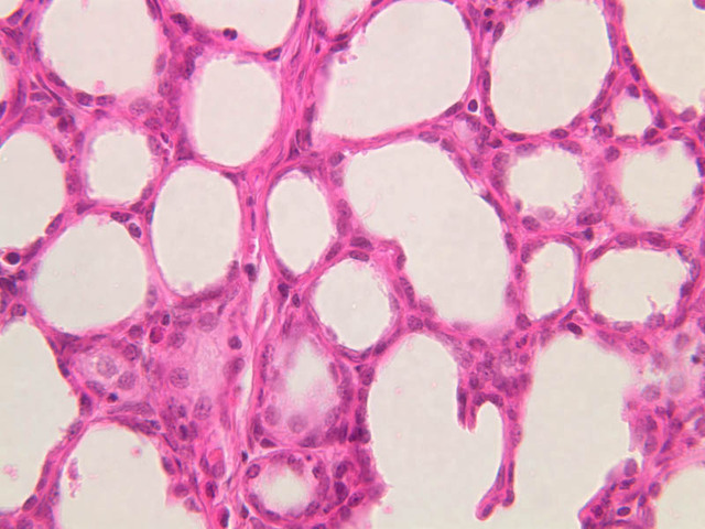
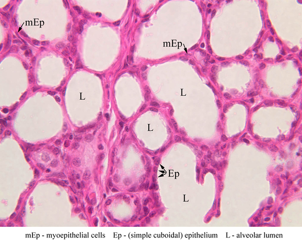
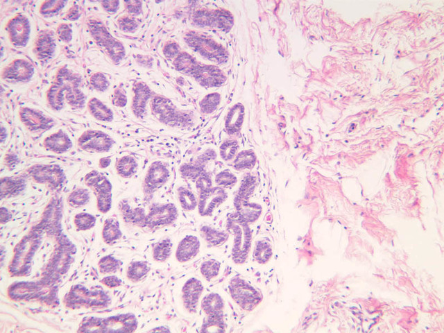
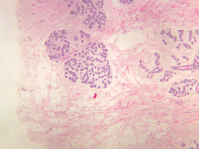
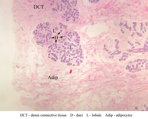
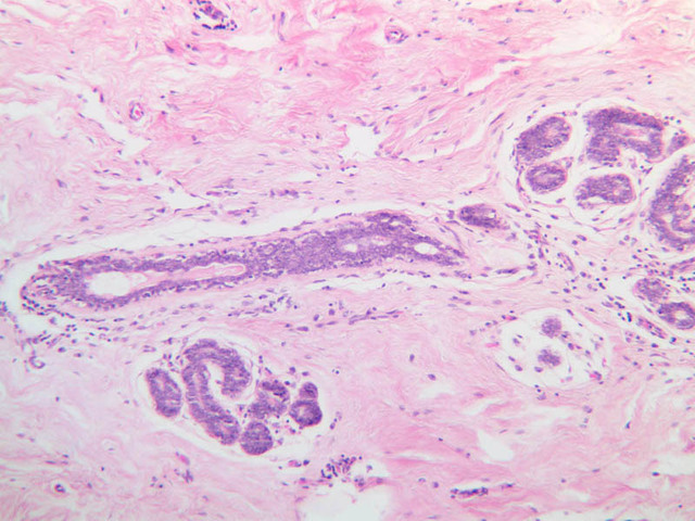
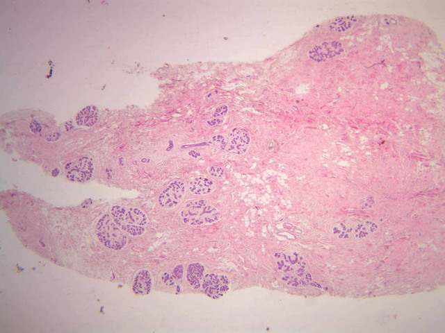
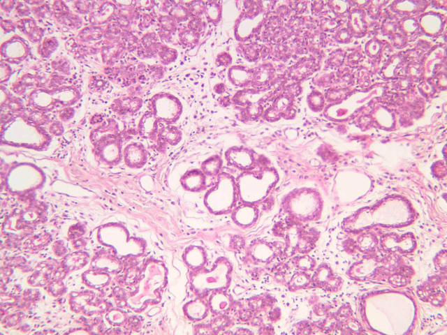
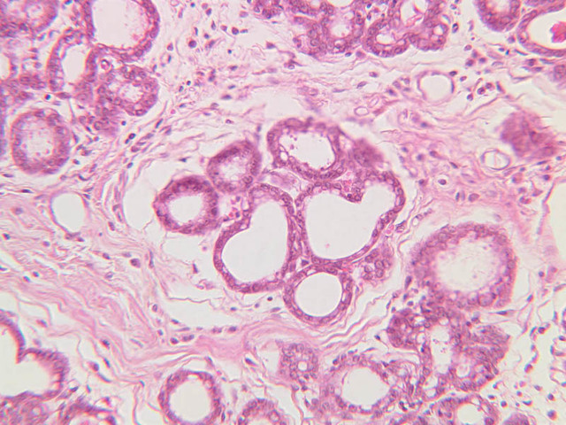
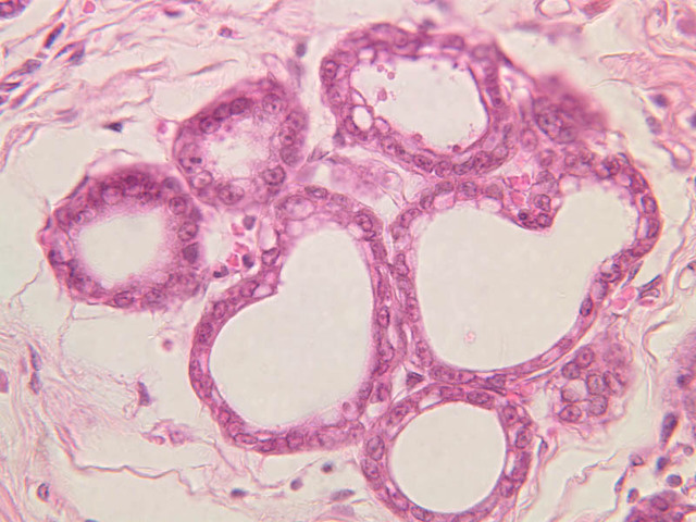
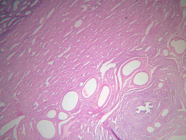
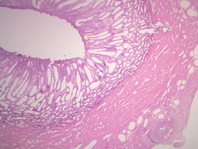
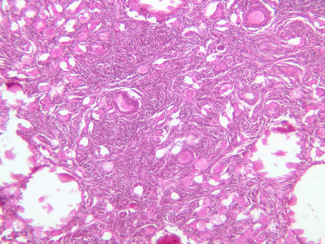
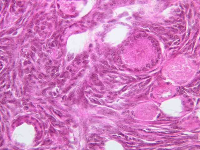
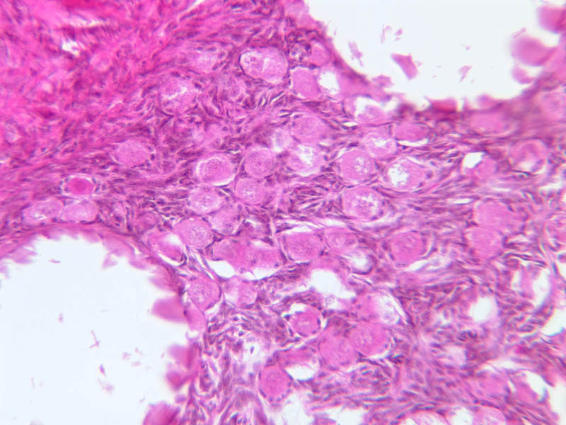
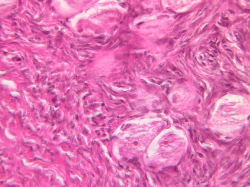
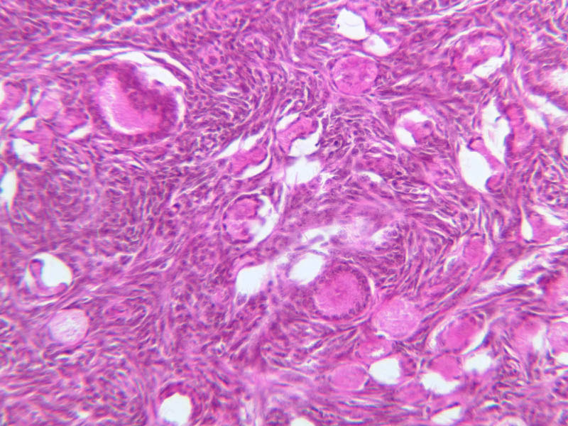
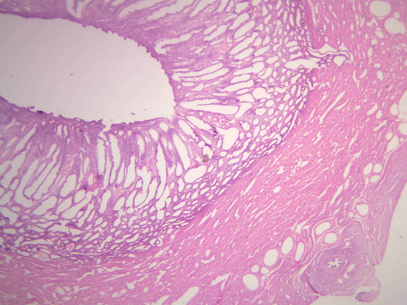
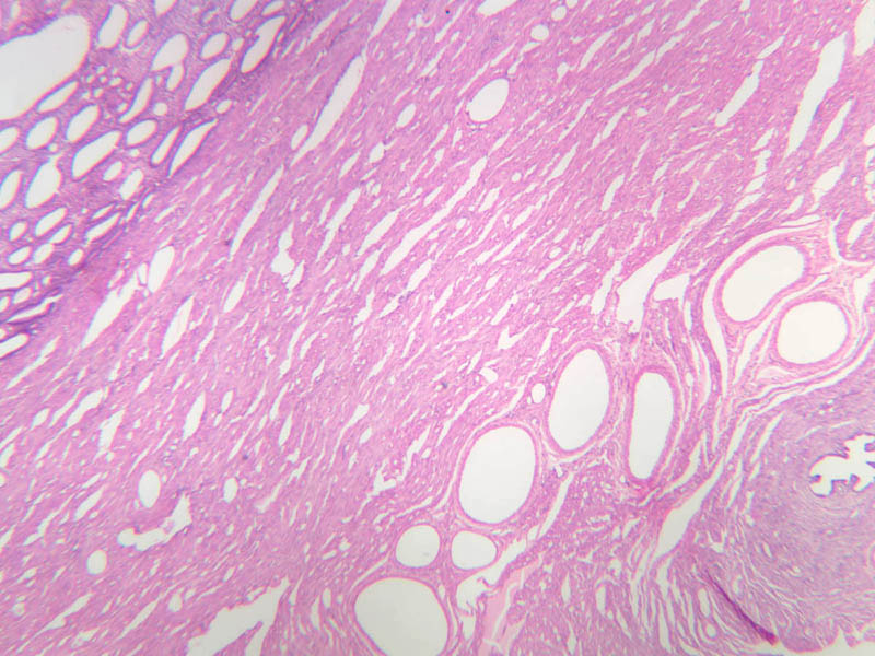
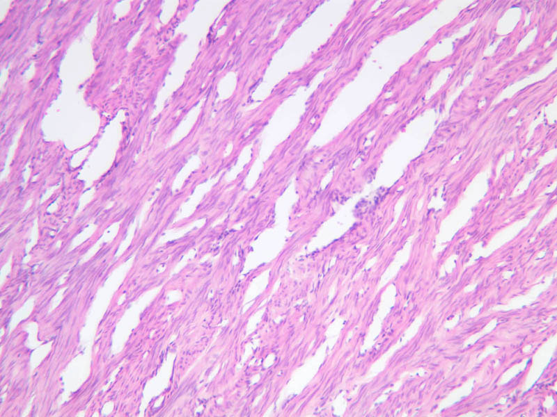
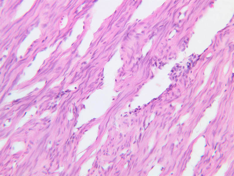
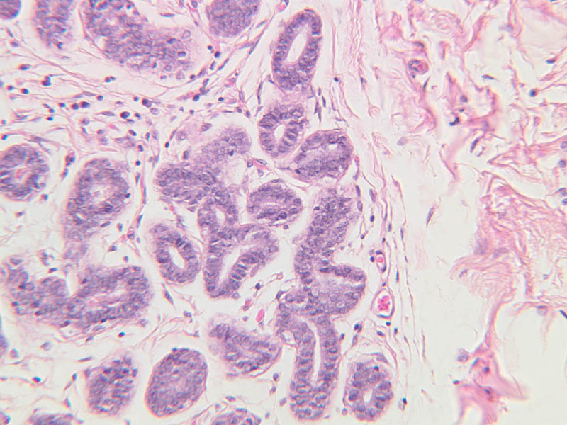
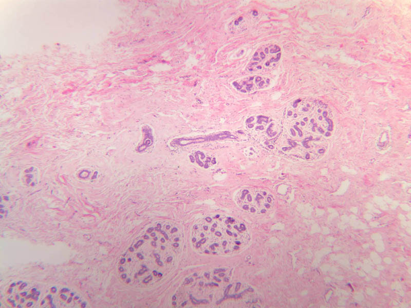
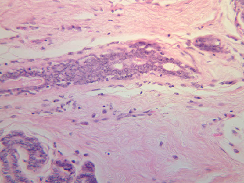
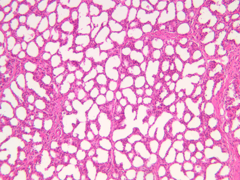
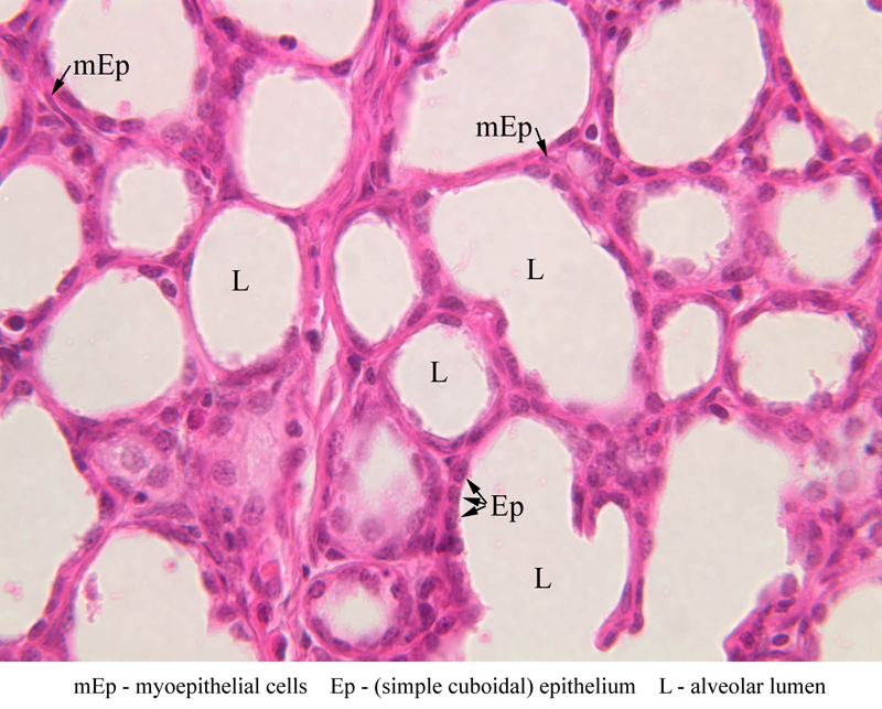
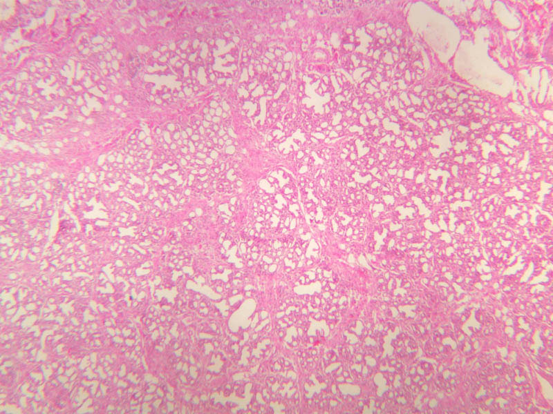
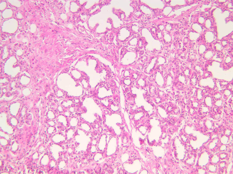
The resting or inactive mammary gland consists of predominantly dense connective tissue with small clusters of ducts and a few glandular elements (slide A-93 [2.5x-labeled, 10x, 20x, 40x] [1x, 2.5x, 10x, 20x]). It is difficult to differentiate between small ducts and alveoli as each is lined by simple low cuboidal cells. Lobes and lobules are not well defined. During puberty, predominantly under the influence of the ovarian hormone estrogen, the glandular or epithelial ducts proliferate and begin to differentiate into clusters of ductal and alveolar units termed terminal ductal lobule units (TDLUs). The non-pregnant gland will form multiple TDLUs that will not fully differentiate until pregnancy-induced growth. TDLUs are classified into Type I, Type II and Type III based on density of the ductules within each lobular unit. Type I and Type II lobules are typical of inactive glands that have not undergone a pregnancy cycle. Type III lobules are seen only in active glands or in inactive glands that have been through a pregnancy. Intralobular connective tissue is loose connective tissue that surrounds the alveoli and ducts within a lobule. The interlobular connective tissue is dense and contains considerable adipose tissue. During pregnancy, predominantly under the influence of the ovarian hormone progesterone and the pituitary hormone prolactin, the glandular elements proliferate and differentiate to form milk-secreting units. In later stages of pregnancy, alveolar development becomes prominent and the amount of connective tissue and adipose tissue decreases. The secretory cells hypertrophy and accumulate secretory product. The mammary gland in its active state is a compound tubuloalveolar gland (slide A-92 [10x, 20x, 40x-labeled] [2.5x, 10x, 20x, 40x]; A-94 [2.5x, 10x, 20x, 40x]). At this time, the gland is predominantly glandular tissue. Each alveolus is lined by a simple cuboidal epithelium. At the base of these cells, and within the alveolar basal lamina, are the stellate-shaped myoepithelial cells that are highly contractile and function to facilitate milk ejection.Mammary Gland Image Gallery


Mammary Gland Table of Identifications
| Row | Structure | Abbreviation | Optimal Stain | Representative Section | Note |
|---|---|---|---|---|---|
| 1 | Dense Connective Tissue | DCT | H&E | |
|
| 2 | Duct | D | H&E | |
|
| 3 | Lobule | L | H&E | |
|
| 4 | Adipocytes | Adip | H&E | |
|
| 5 | Myoepithelial Cells | mEp | H&E | |
|
| 6 | Simple Cuboidal Epithelium | Ep | H&E | |
|
| 7 | Alveolar Lumen | L | H&E | |
Top of page
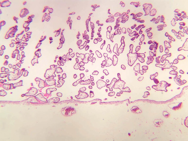
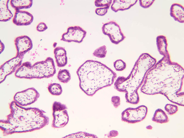
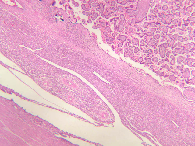
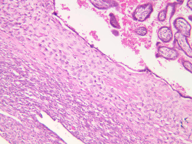
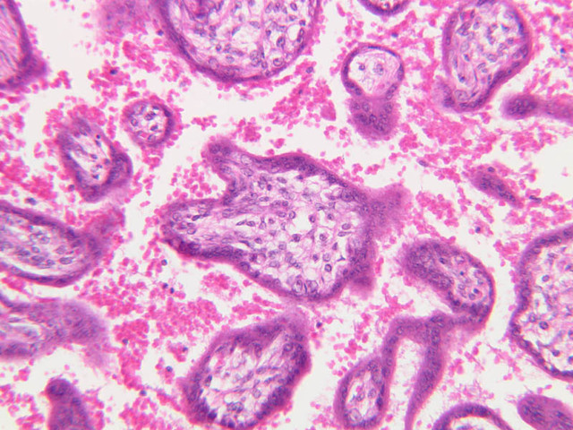
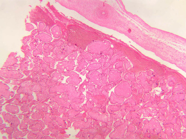
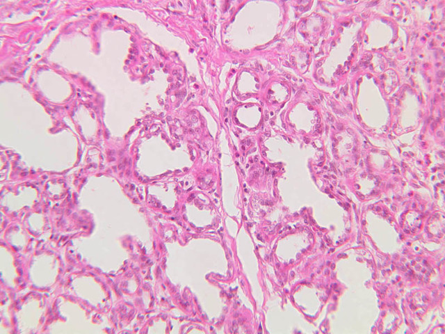
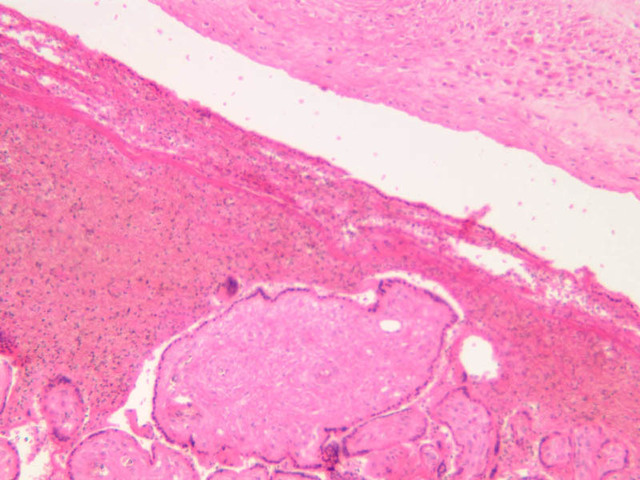
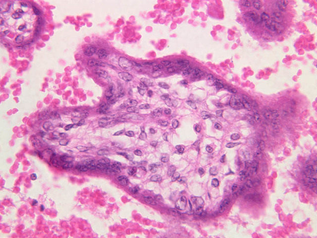
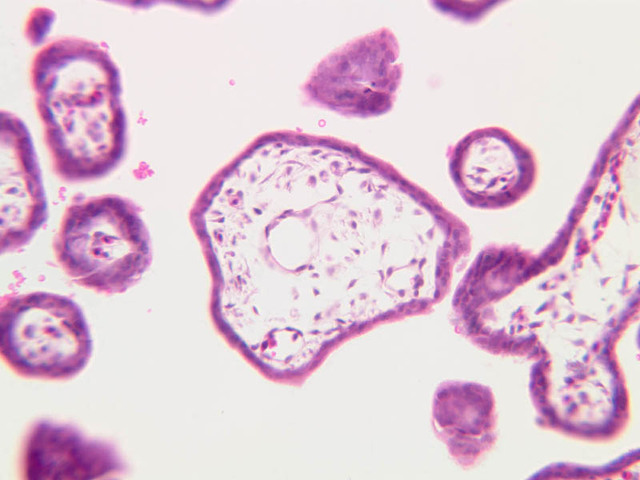
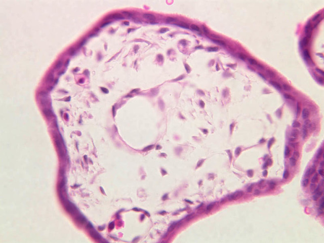
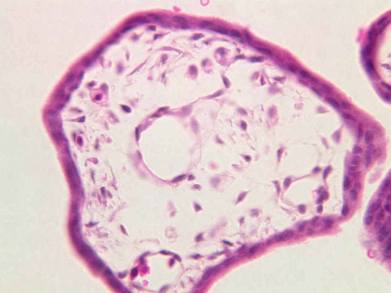
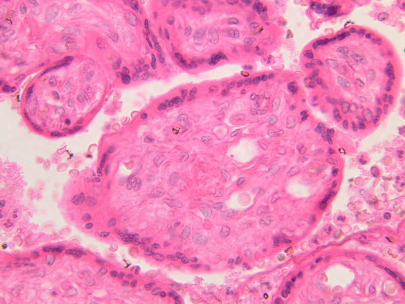
Placenta
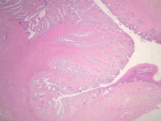
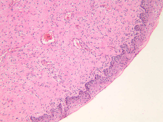
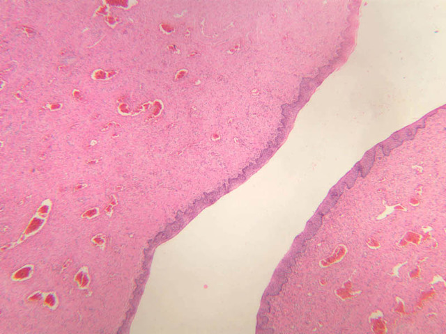
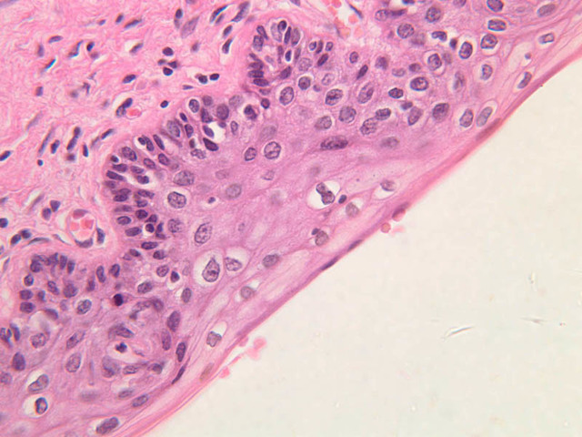
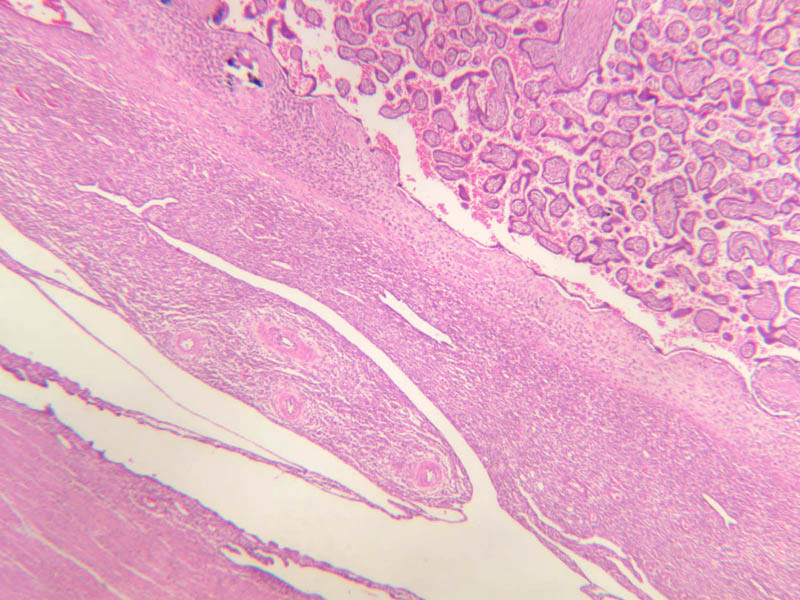
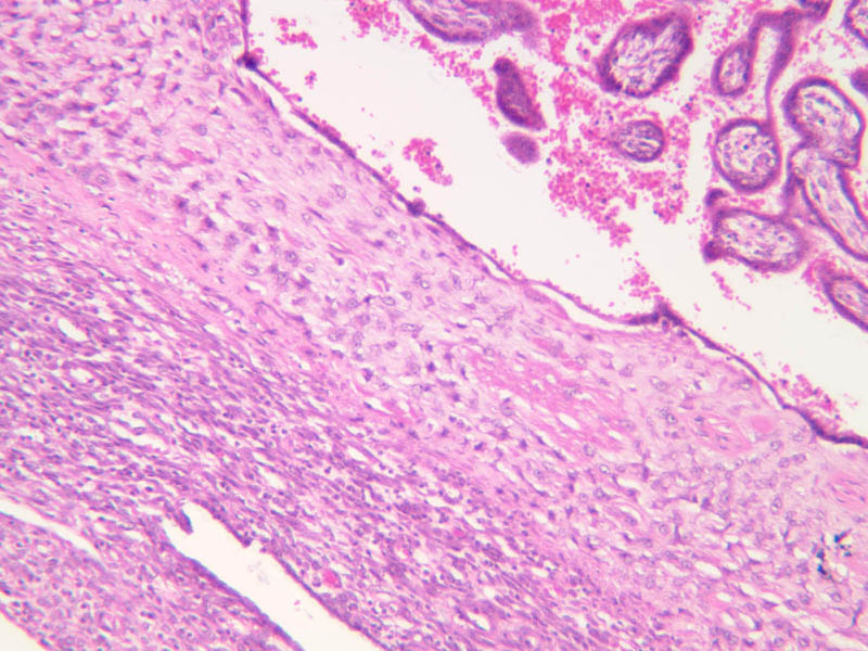
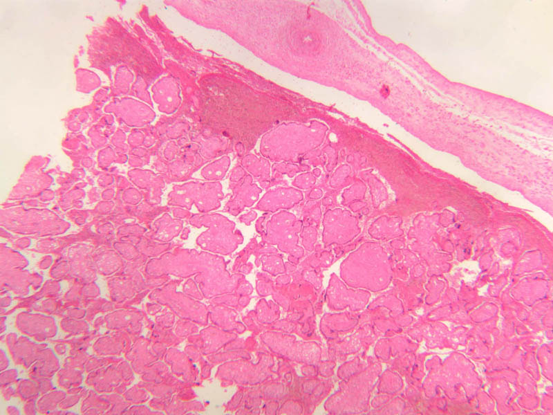
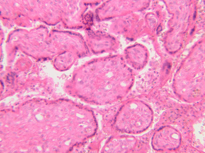
Slide A-98 (monkey placenta [2.5x, 10x, 20x, 40x]) is a first trimester placenta and A-97 (monkey placenta [2.5x, 10x, 20x, 40x]) is from a third trimester pregnancy. What are the differences? Compare to the term human placenta, A-96 ([2.5x, 10x, 20x, 40x]).Placenta Image Gallery


Top of page
Chapter Sixteen Review
Review of Slides


Review of Identifications
| Row | Structure | Abbreviation | Optimal Stain | Representative Section | Note |
|---|---|---|---|---|---|
| 1 | Fimbria | (none) | H&E | |
|
| 2 | Mesovarium | (none) | H&E | |
|
| 3 | Ovary | (none) | H&E | |
|
| 4 | Corpus Luteum | (none) | H&E | |
|
| 5 | Medulla (Ovarian) | (none) | H&E | |
|
| 6 | Cortex (Ovarian) | (none) | H&E | |
|
| 7 | Corpus Albicans | (none) | H&E | |
|
| 8 | Follicles | (none) | H&E | |
|
| 9 | Tunica Albuginea | (none) | H&E | |
|
| 10 | Germinal Epithelium | (none), GE | H&E | |
|
| 11 | Primordial Follicle | (none), Prim | H&E | |
|
| 12 | Follicle Cells | FC | H&E | |
|
| 13 | Nucleus of Oocyte | N | H&E | |
|
| 14 | Early Primary Follicle | (none) | H&E | |
|
| 15 | Granulosa Cells | GC | H&E | |
|
| 16 | Zona Pellucida | ZP | H&E | |
|
| 17 | Basal Lamina | BL | H&E | |
|
| 18 | Theca Folliculi | TF | H&E | |
|
| 19 | Late Primary Follicle | (none) | H&E | |
|
| 20 | Stratum Granulosum | GC | H&E | |
|
| 21 | Theca Interna | TI | H&E | |
|
| 22 | Theca Externa | TE | H&E | |
|
| 23 | Oocyte | O | H&E | |
|
| 24 | Antrum | A | H&E | |
|
| 25 | Primary Follicle | PF | H&E | |
|
| 26 | Secondary Follicle | SF | H&E | |
|
| 27 | Atretic Follicle | AF | H&E | |
|
| 28 | Thecal Cells | TF | H&E | |
|
| 29 | Corona Radiata (future) | CR | H&E | |
|
| 30 | Cumulus Oophorus | CO | H&E | |
|
| 31 | Former Follicular Cavity | (none) | H&E | |
|
| 32 | Granulosa Lutein Cells | GLC | H&E | |
|
| 33 | Theca Lutein Cells | TLC | H&E | |
|
| 34 | Uterine Tube (Interstitial) | (none) | H&E | |
|
| 35 | Uterus | (none) | H&E | |
|
| 36 | Broad Ligament | (none) | H&E | |
|
| 37 | Uterine Tube (Isthmus) | (none) | H&E | |
|
| 38 | Uterine Tube (Infundibulum) | (none) | H&E | |
|
| 39 | Uterine Tube (Ampulla) | (none) | H&E | |
|
| 40 | Serosa | (none) | H&E | |
|
| 41 | Muscularis | (none) | H&E | |
|
| 42 | Mucosa | (none) | H&E | |
|
| 43 | Nonciliated Peg Cells | (none) | H&E | |
|
| 44 | Ciliated Cells | (none) | H&E | |
|
| 45 | Perimetrium | (none) | H&E | |
|
| 46 | Myometrium | (none) | H&E | |
|
| 47 | Endometrium | (none) | H&E | |
|
| 48 | Uterine Lumen | (none) | H&E | |
|
| 49 | Blood Vessels | (none) | H&E | |
|
| 50 | Cervical Glands | (none) | H&E | |
|
| 51 | Cervix | (none) | H&E | |
|
| 52 | Vagina | (none) | H&E | |
|
| 53 | Stratified Squamous Epithelium | (none) | H&E | |
|
| 54 | Lymphatic Tissue | (none) | H&E | |
|
| 55 | Dense Connective Tissue | DCT | H&E | |
|
| 56 | Duct | D | H&E | |
|
| 57 | Lobule | L | H&E | |
|
| 58 | Adipocytes | Adip | H&E | |
|
| 59 | Myoepithelial Cells | mEp | H&E | |
|
| 60 | Simple Cuboidal Epithelium | Ep | H&E | |
|
| 61 | Alveolar Lumen | L | H&E | |
Top of page
Comments
- That's funny. The comments just happen that way when I upload from a batch file (or I can specify a name that will be applied universally to all extracted images). I meant the file names... they were incorrectly labeled based on the text, and the text has references to slides that don't exist (image-wise). I'm just having to read through the text and glance at the slides and figure out which images need to go where. I'll be doing a lot of editing when I link the text to the images in a few minutes. -- AshleyLPistorio - 10 Jul 2007
- Yea, I noticed the redundancy "extracted from zip." I don't think so. I have not heated the oven in 10 years. Refuse to clean it. -- LorenEvey - 10 Jul 2007
- Wow. Ever wonder if you spend too much time in your kitchen? I'll be attacking the uterus now. I still have to figure out which pictures go in what order... they're all named rather redundantly.
 -- AshleyLPistorio - 10 Jul 2007
- Let's get going with this uterus. I want to see the uterovaginal autonomic plexus receiving fibers from the hypogastric nn. and the pelvic splanchnics. At last! -- LorenEvey - 10 Jul 2007
Edit | Attach | Print version | History: r2 < r1 | Backlinks | View wiki text | More topic actions
Topic revision: r2 - 20 Jun 2015, LorenEvey

- Epithelium
- Connective Tissue
- Muscle
- Nervous Tissue
- Cardiovascular System
- Skin Appendages and Sensory Receptors
- Lymphatic System
- Cartilage and Bone
- Respiratory System
- Peripheral Blood and Bone Marrow
- Oral Cavity and Salivary Glands
- Esophagus and Gastrointestinal Tract
- Pancreas, Liver, and Gall Bladder
- Endocrine Organs
- Male Reproductive System
- Female Reproductive System
- Urinary System
Ideas, requests, problems regarding Medical Histology? Send feedback