 |
|
You are here: Medical Histology>Main Web>AtlasContents>RespiratorySystemAtlas09 (20 Jun 2015, LorenEvey)Edit Attach
Chapter Nine: Respiratory System


Introduction
In studying the microscopic anatomy of the respiratory system keep in mind that inspired air must be brought to the right temperature and humidity, and be filtered of particulate matter. Expired air must not be so humid that the body loses excessive moisture. Since the introduction of air into the moist body spaces of airways and lungs provides a means for the invasion of bacteria and viruses, the body must have protective devices to counteract the danger. Finally, the principal purpose of air transfer to and from the blood must be satisfied. Structures adapted for the accomplishment of these several purposes together comprise the respiratory tree. Consequently, the respiratory tree is divided into: 1) the portions which conduct, filter, and condition the air, and 2) those portions which are the actual sites of gaseous exchange with the blood. The innumerable pulmonary alveoli, blind but interconnected endings of the tree, comprise the exchange sites; the remainder of the airways accomplish the other purposes. Prior to learning the microarchitecture of the Respiratory System, use the table below to review some of the gross anatomy of these tissues:| Structure | Image |
|---|---|
| Internal Anatomy of the Nose | |
| Anatomy of the Larynx | |
| Internal View of Larynx (through airway) | |
| The Larynx, Trachea, and Bronchi | |
| Gross Anatomical Arrangement of the Lungs | |
| Internal Anatomy of the Lungs | |
Nose
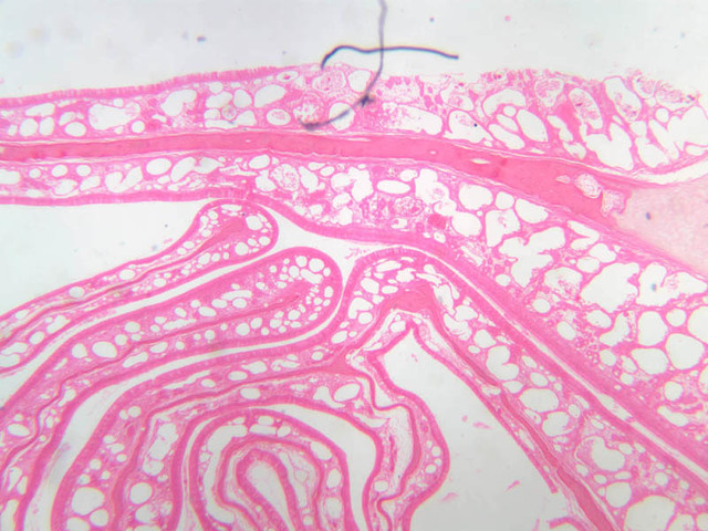
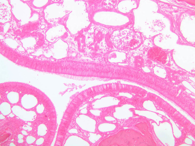
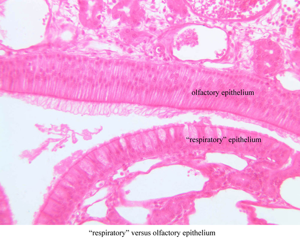
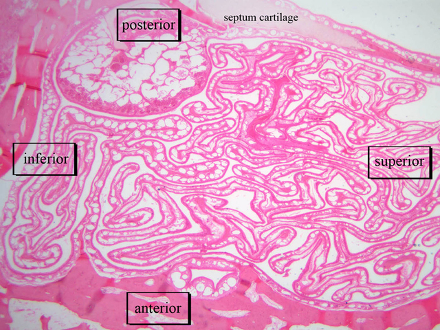
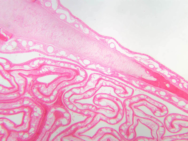
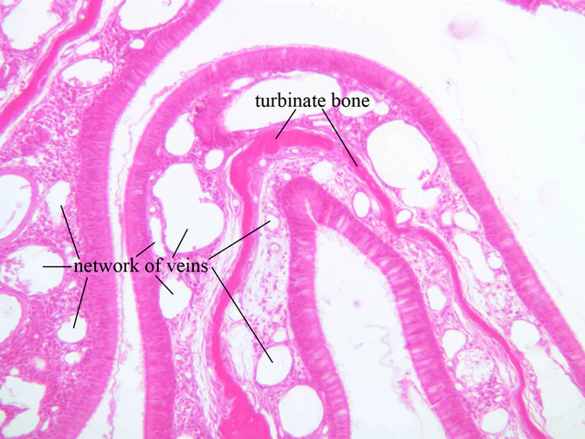
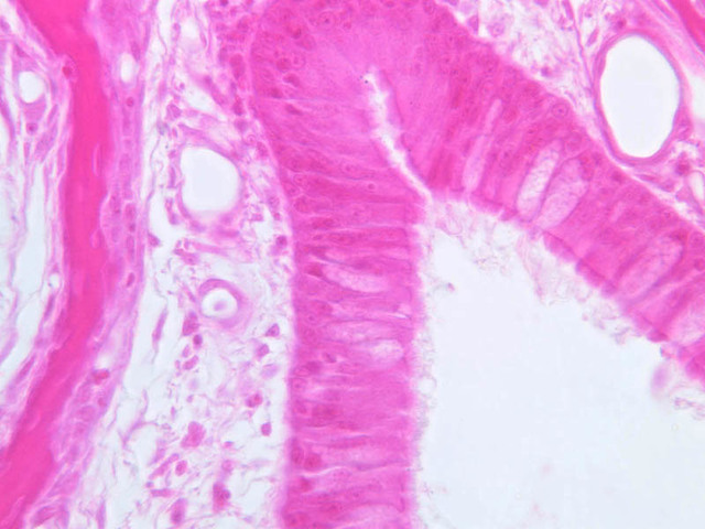
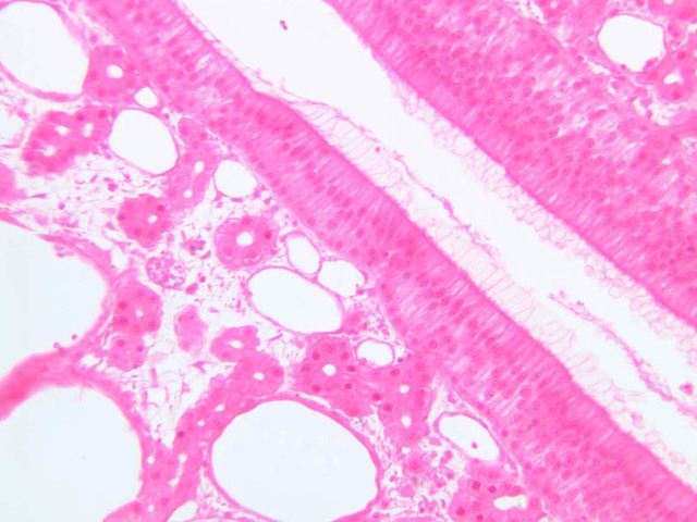
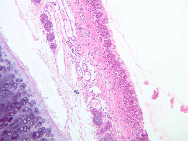
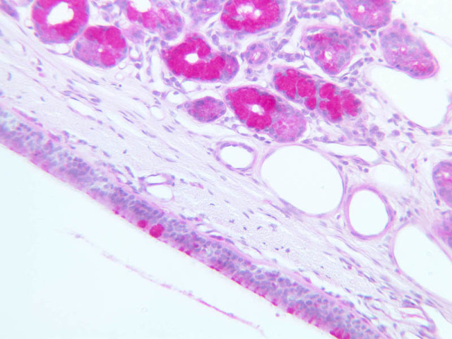
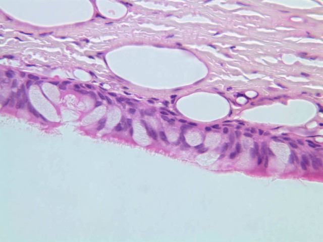
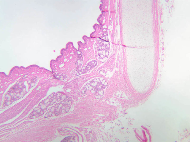
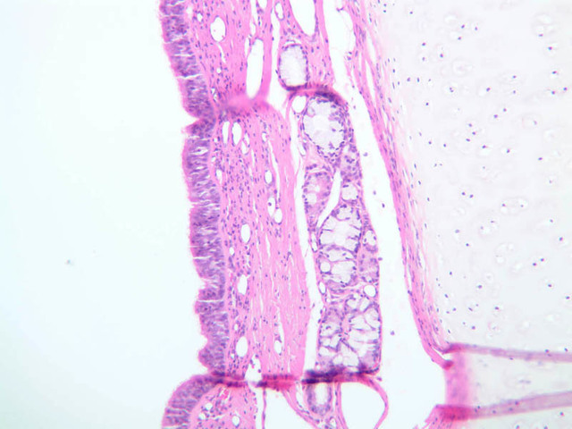
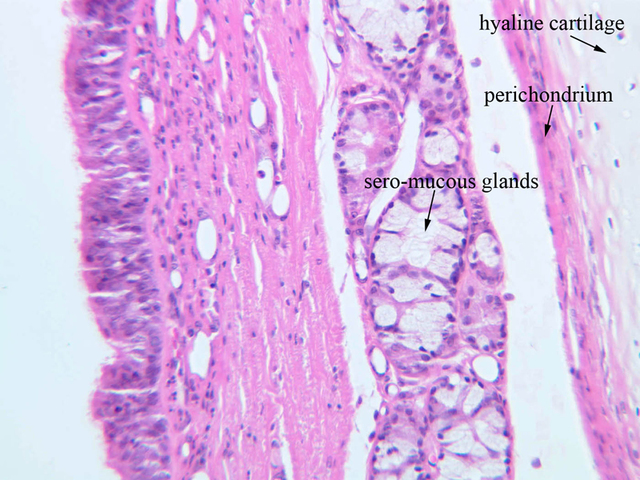
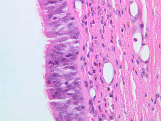
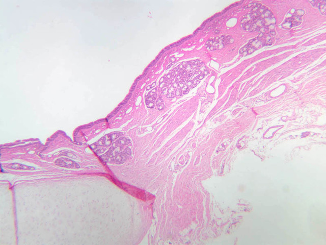
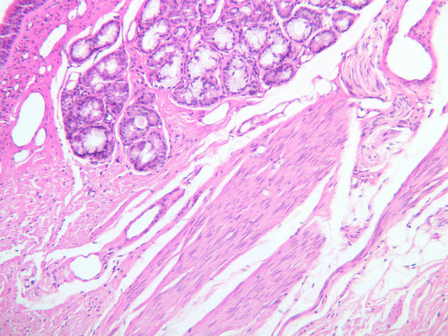
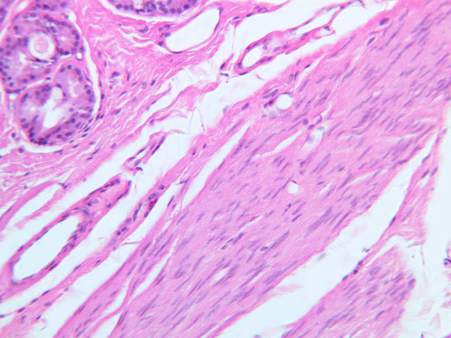
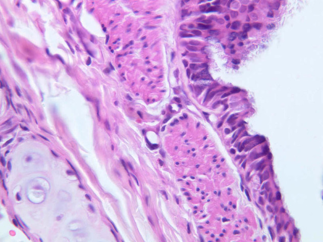
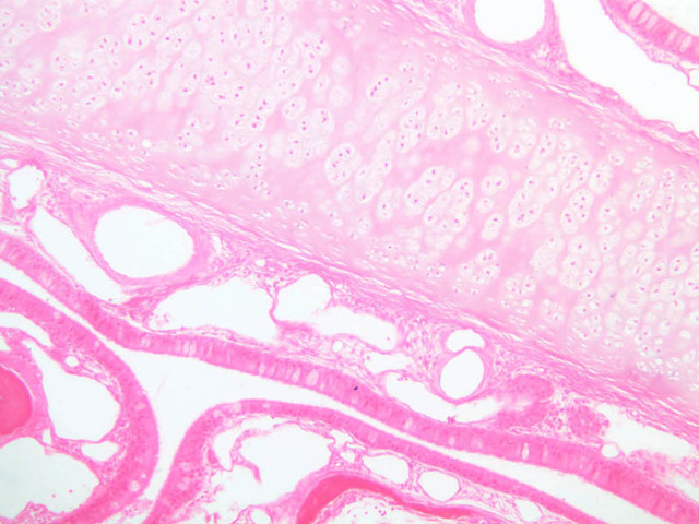
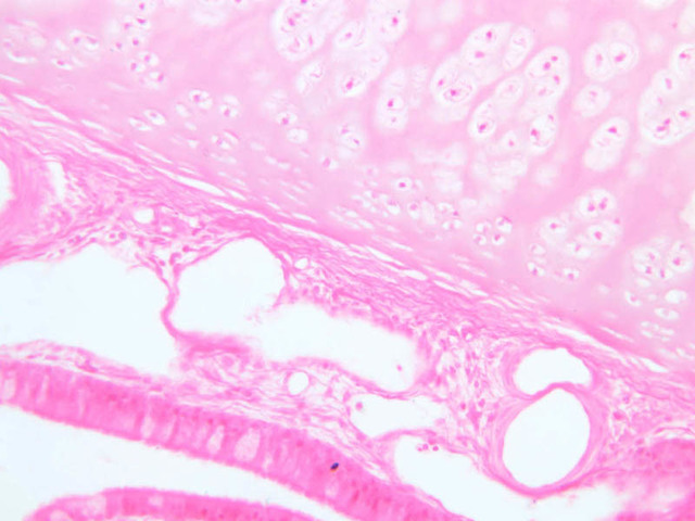
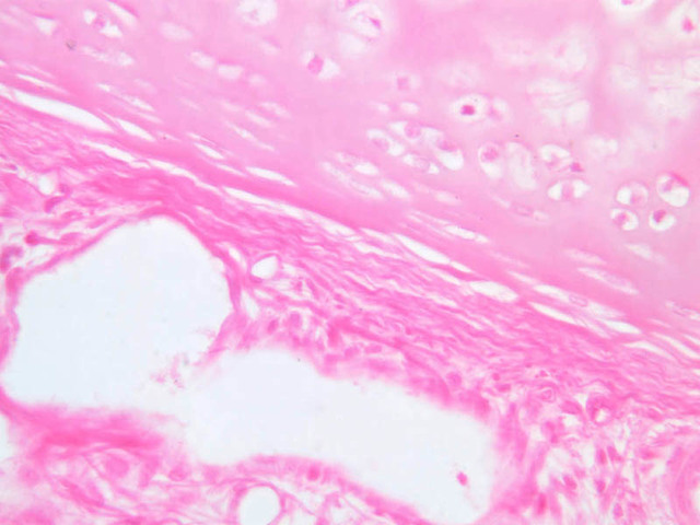
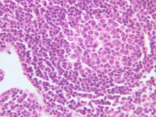
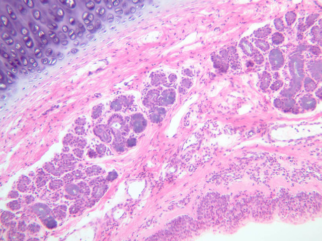
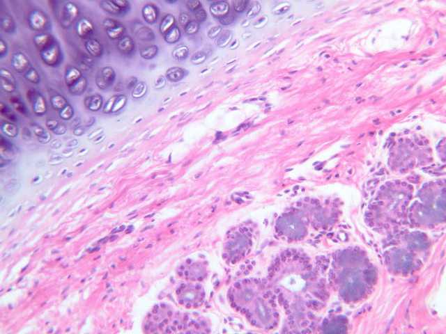
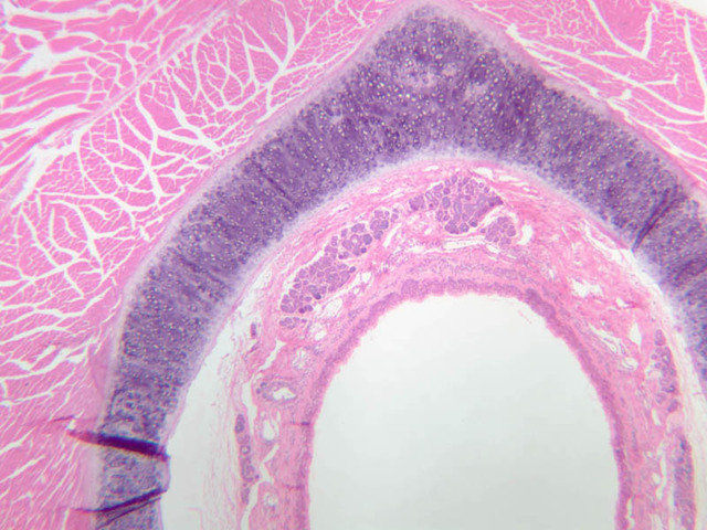
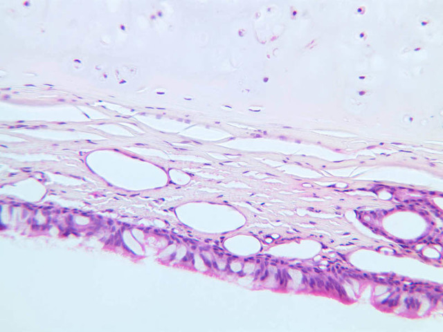
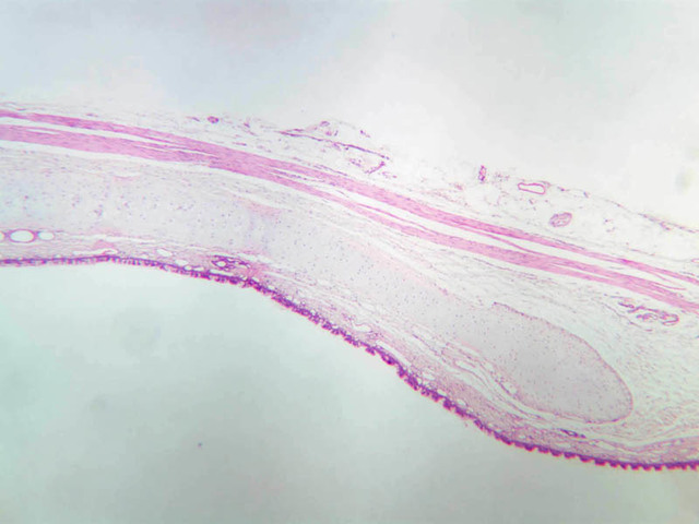
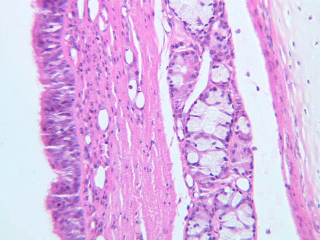
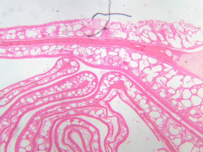
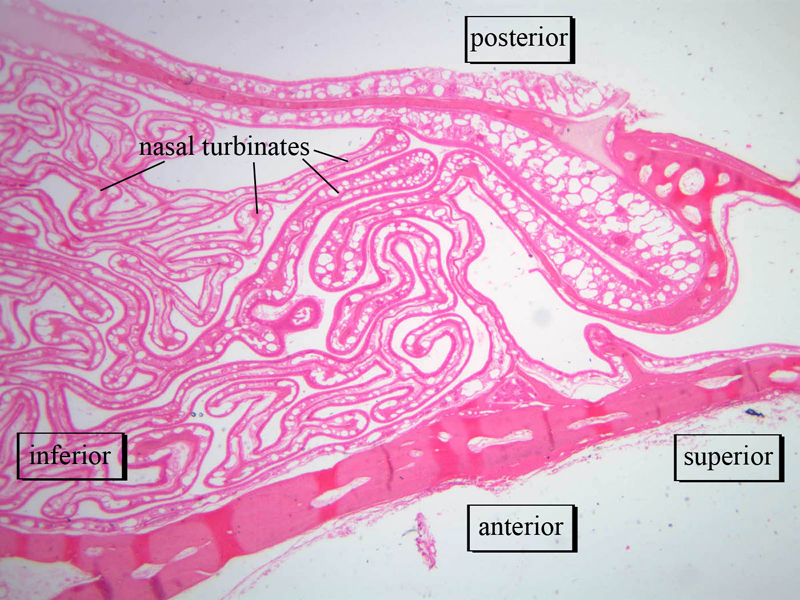
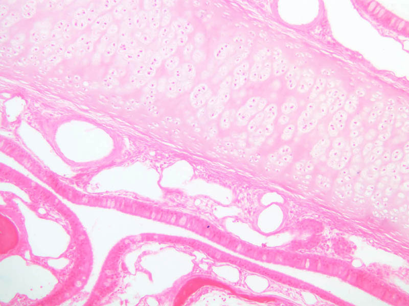
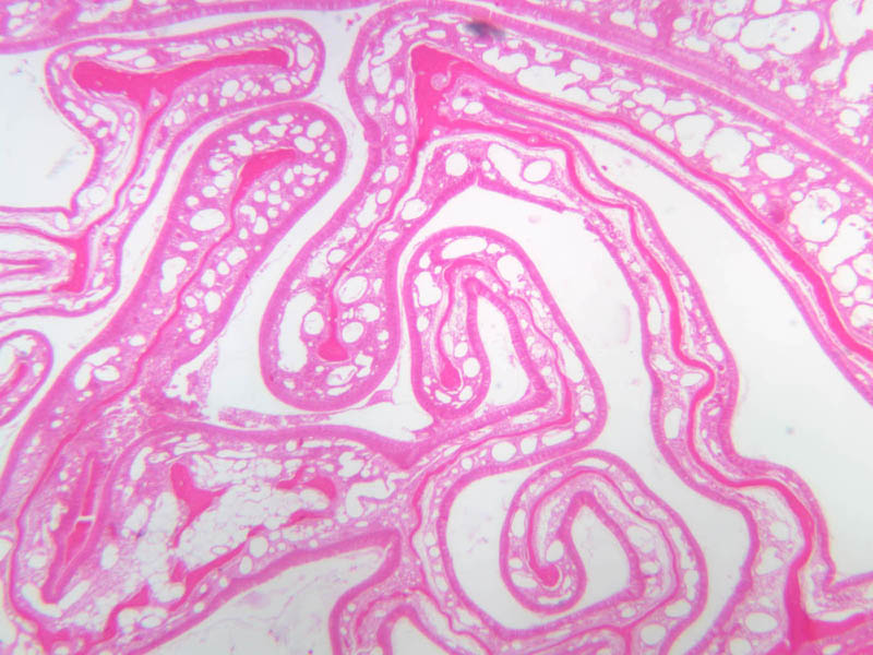
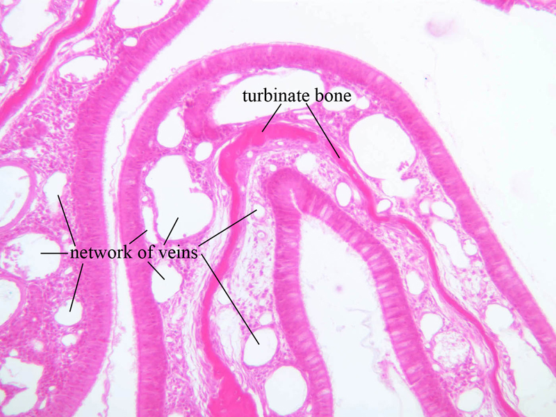
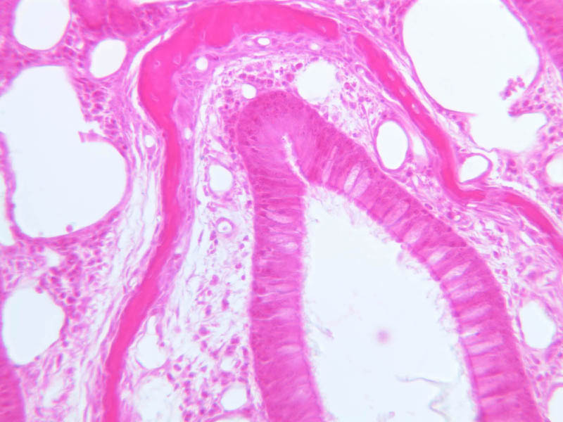
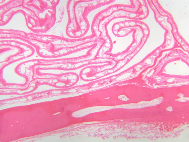
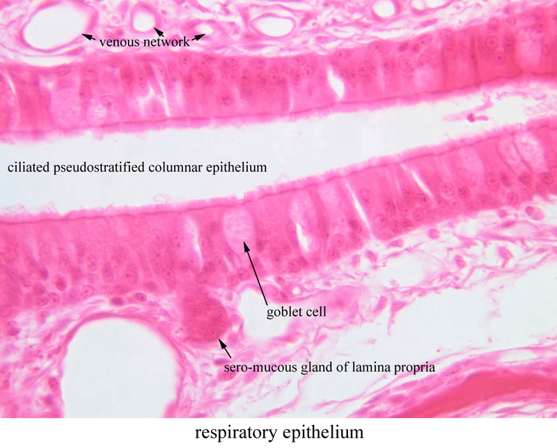
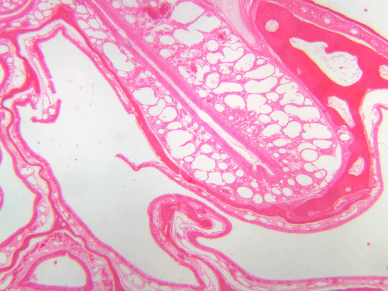
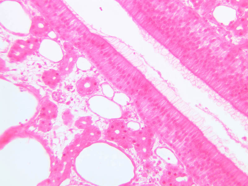
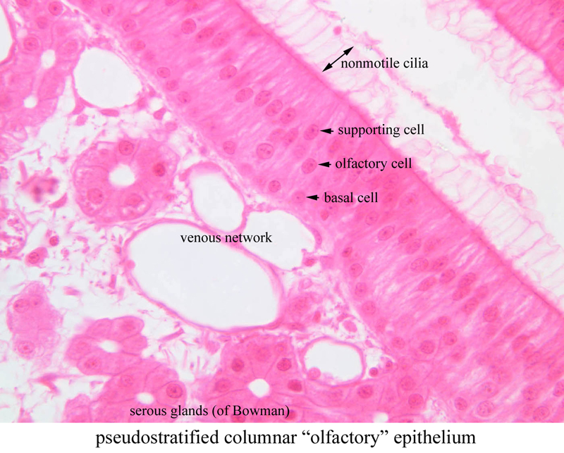
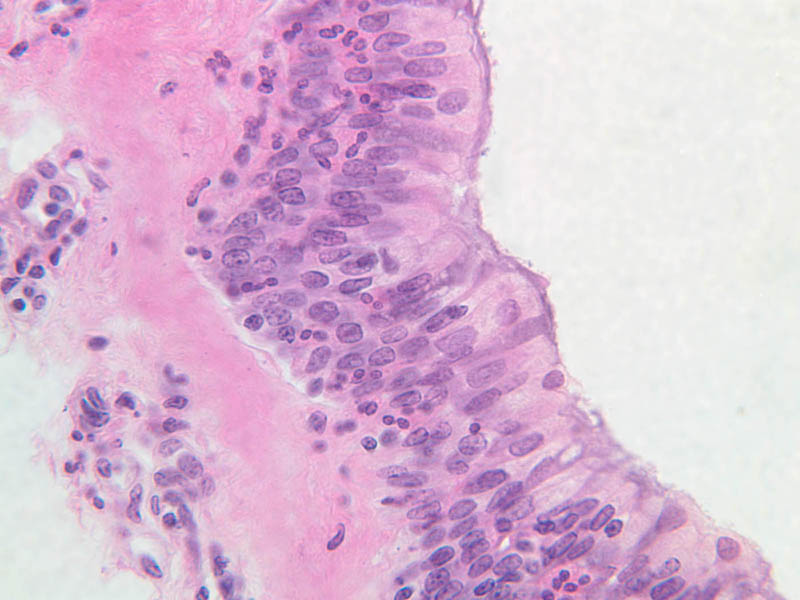
There are two fundamentally different types of epithelium in the nasal cavity, which will be discussed here (A-70 [2.5x, 10x, 20x-labeled, 40x]). Initial filtration, warming and moistening of inspired air occurs in the nose (A-70, nasal cavity, cat [1x-labeled, 1x-labeled]). Coarse hairs that are present in the nasal vestibule screen large particulate matter from the inspired air. As the hairs thin out internally, the nasal passages expand into what are called the nasal mucosa of the nose (slide A-70 [2.5x, 10x, 20x, 40x]). This is lined with ciliated pseudostratified columnar epithelium, which also lines most of the conducting passages of the respiratory system and is frequently known as "respiratory epithelium" ([2.5x, 10x-labeled, 20x, 40x]). Associated with this section of the nose are numerous sero-mucous glands located in the lamina propria of the mucosa [2.5x, 10x, 20x, 40x-labeled]). They aid the goblet cells found in the epithelium itself in keeping both the membrane and the air moist. A prominent additional feature of the lamina propria is the extensive network of veins from which heat radiates to warm the passing air. The lamina propria's connective tissue merges with the connective tissue (periosteum) of the nasal skeleton ([2.5x, 10x, 20x, 40x]). The olfactory epithelium is found in the uppermost and posterior portion of the nasal cavity and is normally covered by a layer of secreted material, which is described below. Slide A-70 was prepared from an animal with a complex system of nasal turbinate bones. Look for an area with a very thick epithelium devoid of goblet cells ([2.5x, 10x, 20x, 40x] ([2.5x, 10x, 20x, 40x-labeled]). This epithelium is a pseudostratified columnar type with three cell types: supporting cells, basal cells, and olfactory cells. The olfactory cells are bipolar neurons. The apical surface has a bulbous projection with olfactory cilia which are responsible for sensory transduction of odorous substance. A profusion of subepithelial serous glands (of Bowman) produce a secretion that helps keep the olfactory mucosa moist and serve as a solvent for odiferous substances. Functional terminations of the nerve cells are too small to study adequately with the light microscope, but many examples of small nerve trunks can be seen beneath the epithelium. These trunks are branches of the olfactory nerve and consist of unmyelinated fibers.Nose Image Gallery


Nose Table of Identifications
| Row | Structure | Abbreviation | Optimal Stain | Representative Section | Note |
|---|---|---|---|---|---|
| 1 | Olfactory Epithelium | (none) | H&E | |
|
| 2 | Respiratory Epithelium | (none) | H&E | |
|
| 3 | Turbinate Bone | (none) | H&E | |
|
| 4 | Network of Veins | (none) | H&E | |
|
| 5 | Nonmotile Cell | (none) | H&E | |
|
| 6 | Supporting Cell | (none) | H&E | |
|
| 7 | Olfactory Cell | (none) | H&E | |
|
| 8 | Basal Cell | (none) | H&E | |
|
| 9 | Serous Gland (of Bowman) | (none) | H&E | |
|
| 10 | Pseudostratified Columnar (Olfactory) Epithelium | (none) | H&E | |
|
| 11 | Ciliated Pseudostratified Columnar (Repiratory) Epithelium | (none) | H&E | |
|
| 12 | Goblet Cell | (none) | H&E | |
|
| 13 | Seromucous Gland of Lamina Propria | (none) | H&E | |
Top of page
Larynx
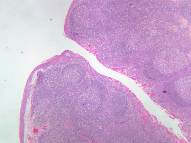
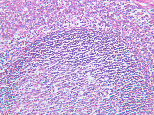
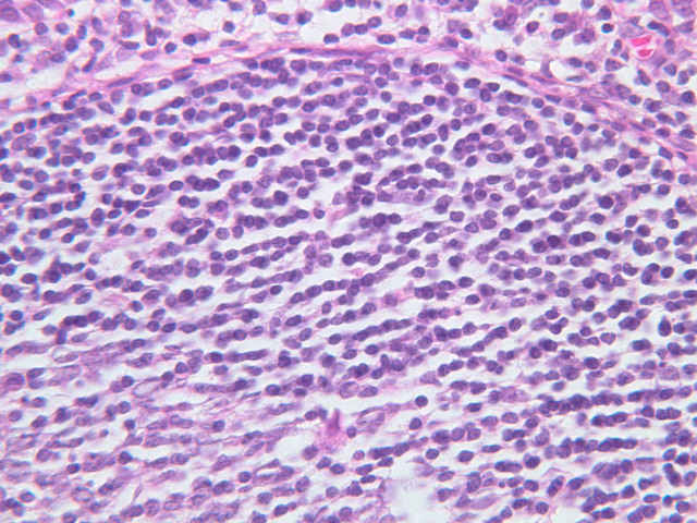
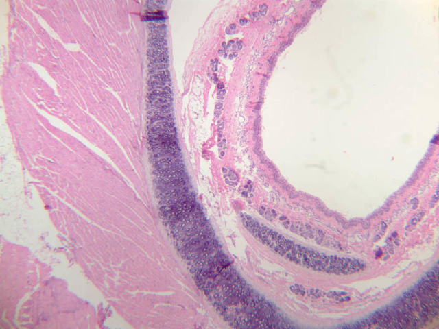
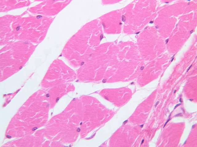
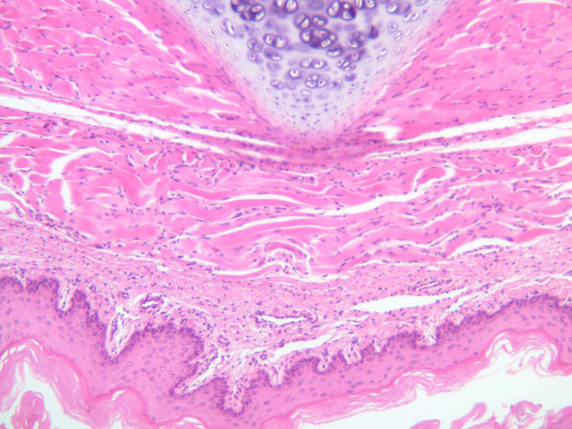
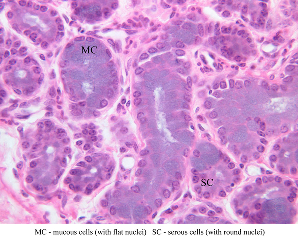
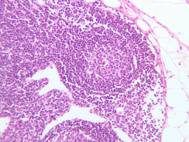
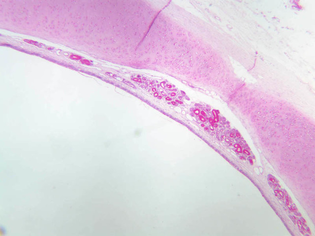
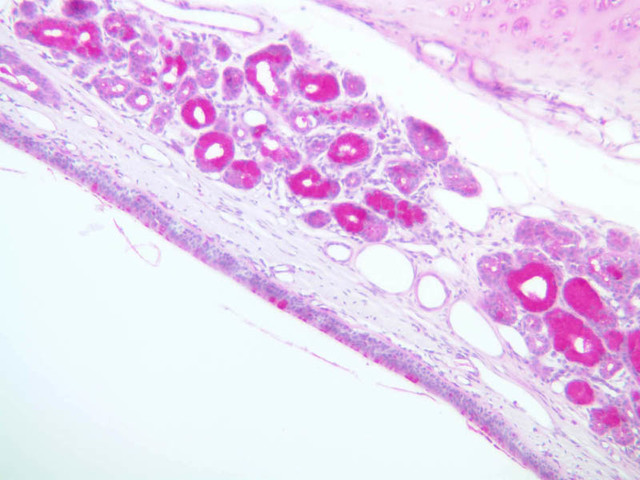
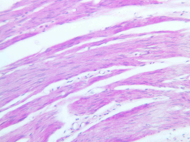
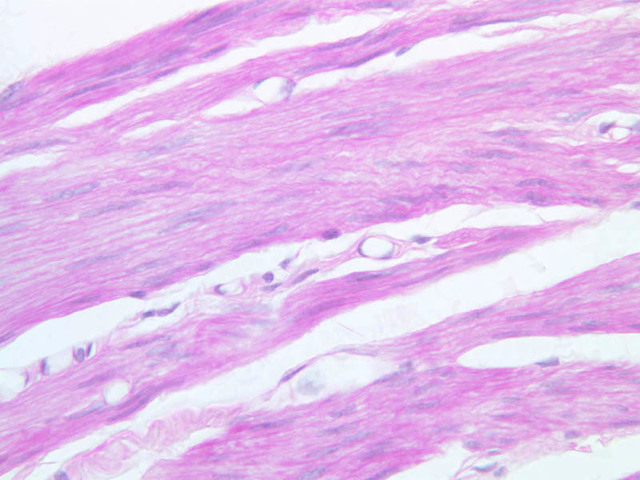
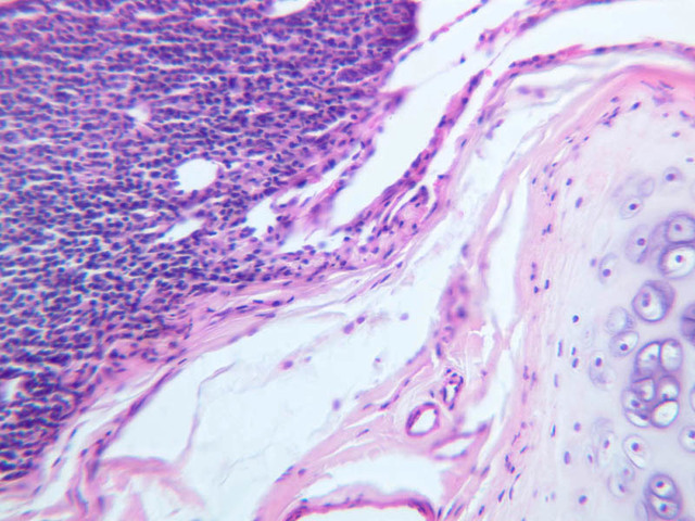
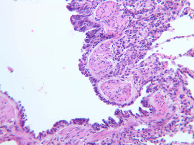
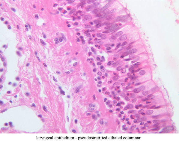
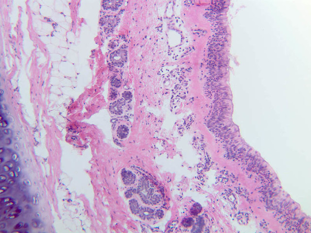
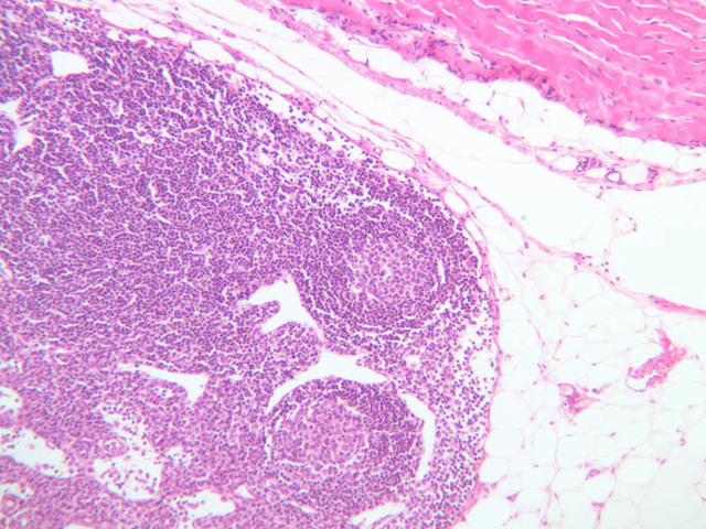
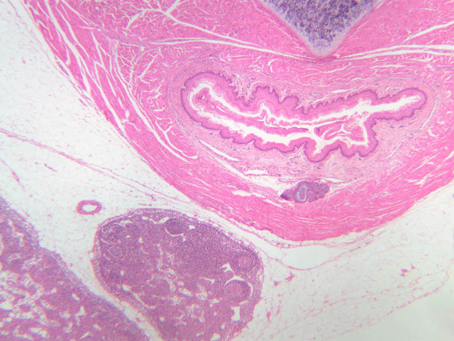
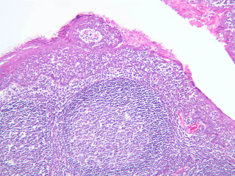
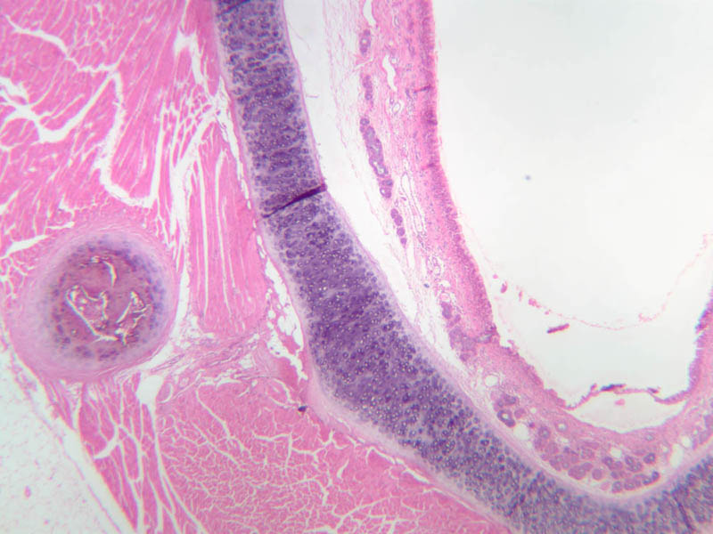
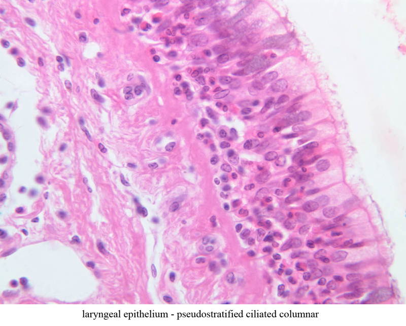
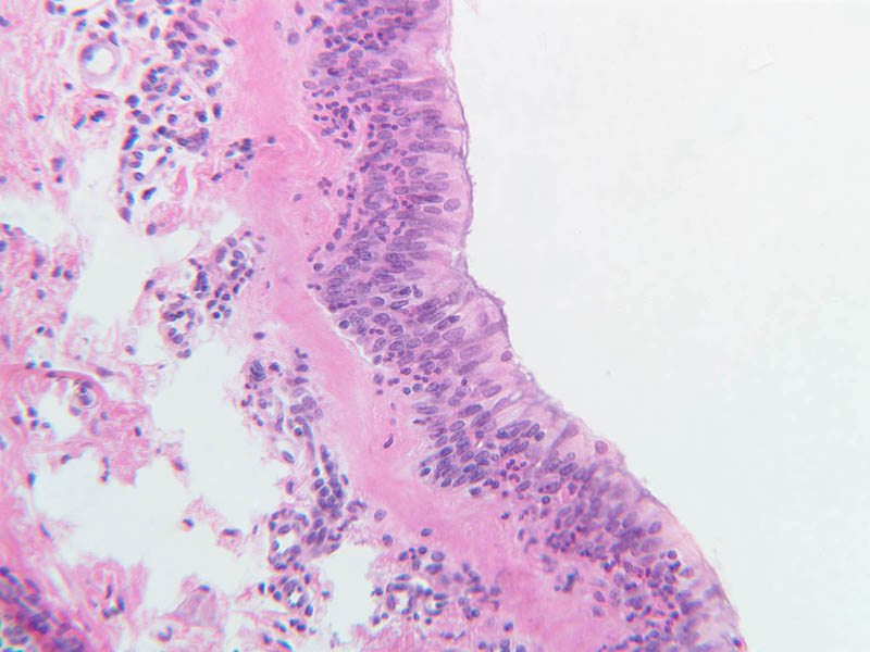
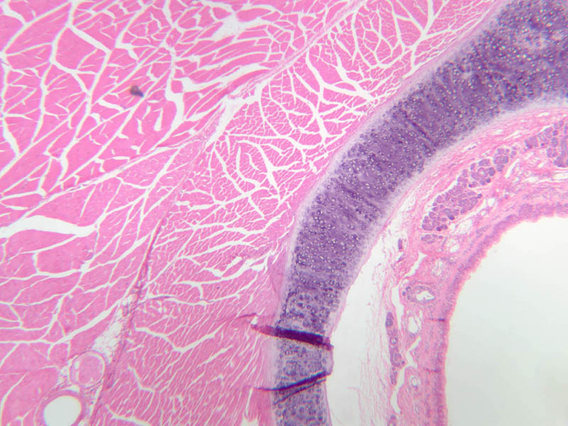
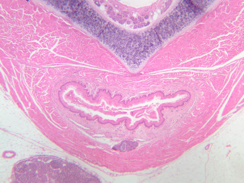
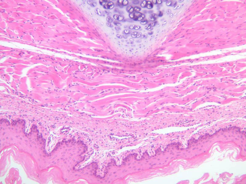
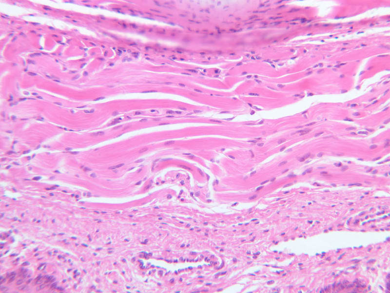
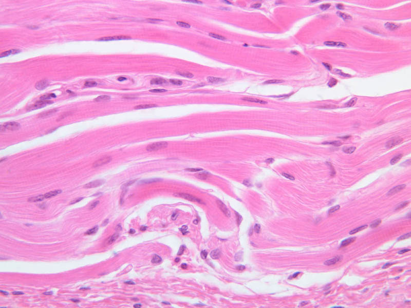
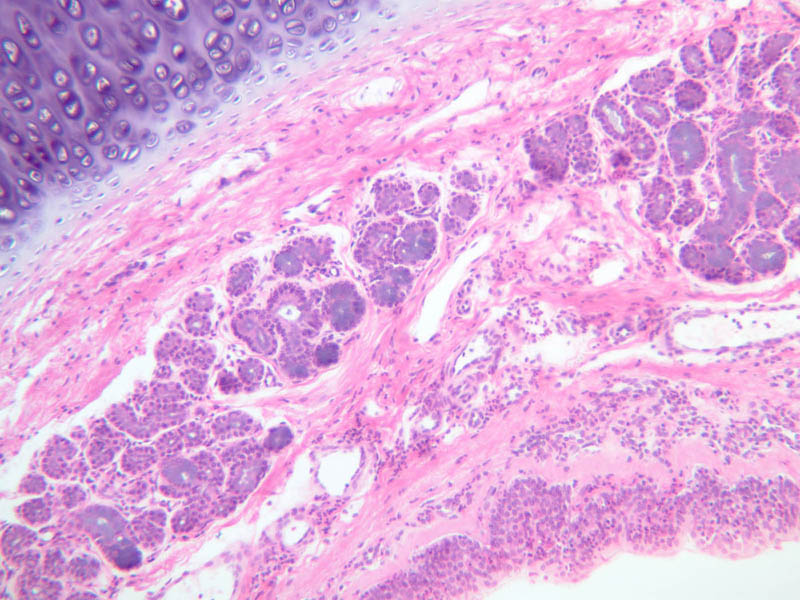
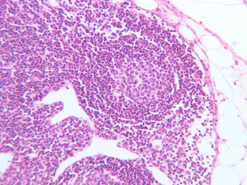
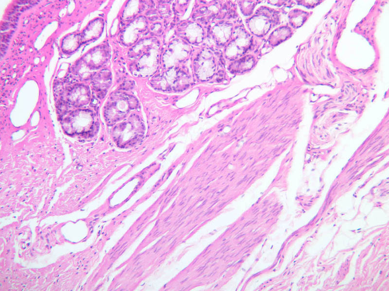
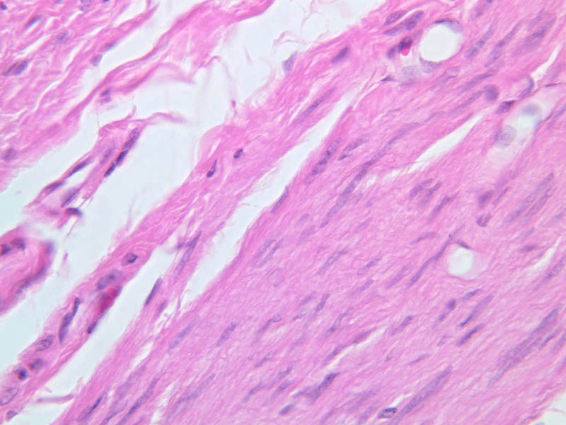
Air passes from the nose to the larynx and trachea. It crosses the alimentary canal through the pharynx. This intersection is ringed by the various clusters of lymphoid tissue called tonsils (slide A-41 [2.5x, 10x, 20x, 40x]). Tonsils are discussed in the lymphatic system laboratory. The location of these bodies has obvious significance for exposing macrophages and immunologically competent cells to foreign organisms. The mucosa of the larynx is continuous with and connects the pharynx and the trachea. Stratified squamous (nonkeratinizing type) epithelium covers the epiglottis and upper half of the larynx and areas subject to wear like the vocal folds. After a transition zone (of stratified columnar epithelium) the remainder of the laryngeal epithelium is pseudostratified ciliated columnar (A-73 [2.5x, 10x, 20x, 40x-labeled] [2.5x, 10x, 20x, 40x]). What type of epithelium do you see in your slide? The larynx (A-73 [2.5x, 10x, 20x, 40x] [2.5x, 10x, 20x, 40x]) is kept open by a series of cartilaginous rings. Characterize the cartilage in the walls as to type and extent. The particular importance of the larynx is clearly demonstrated in its use for the generation of sounds. The lining epithelia, the fibrous elements of the vocal cords and muscles attached to the cords are adapted for this purpose. The respiratory epithelium of upper and lower larynx is replaced over the vocal cords by a thin stratified squamous epithelium. The body of the cords consists of skeletal muscle and elastic connective tissue. Numerous examples of both serous and mucous glands can be seen everywhere beneath the laryngeal epithelium except in the vocal cords themselves ([2.5x, 10x, 20x, 40x-labeled]). Occasional lymphoid nodules may also occur ([2.5x, 10x, 20x, 40x]).Larynx Image Gallery


Larynx Table of Identifications
| Row | Structure | Abbreviation | Optimal Stain | Representative Section | Note |
|---|---|---|---|---|---|
| 1 | Laryngeal Epithelium (Ciliated Pseudostratified Columnar) | (none) | H&E | |
|
| 2 | Mucous Cells (With Flat Nuclei) | MC | H&E | |
|
| 3 | Serous Cells (With Round Nuclei | SC | H&E | |
Top of page
Trachea
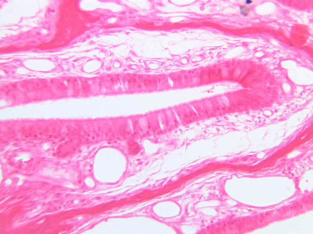
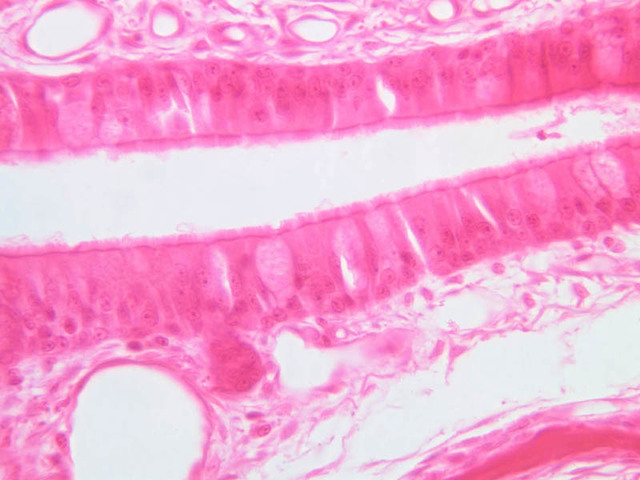
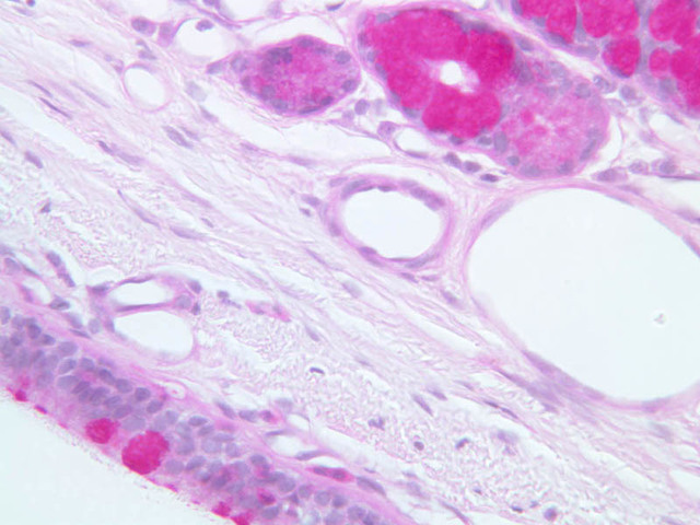
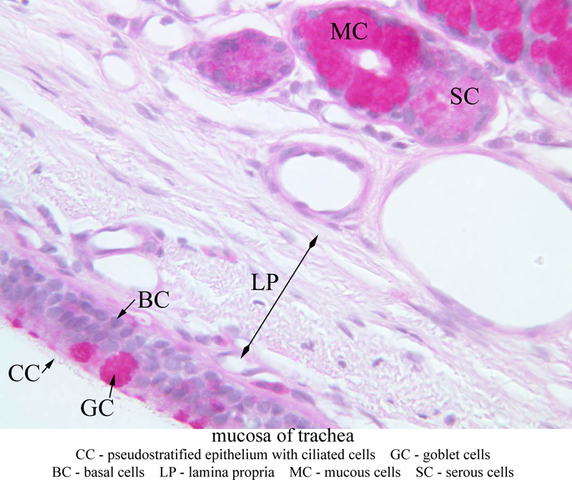
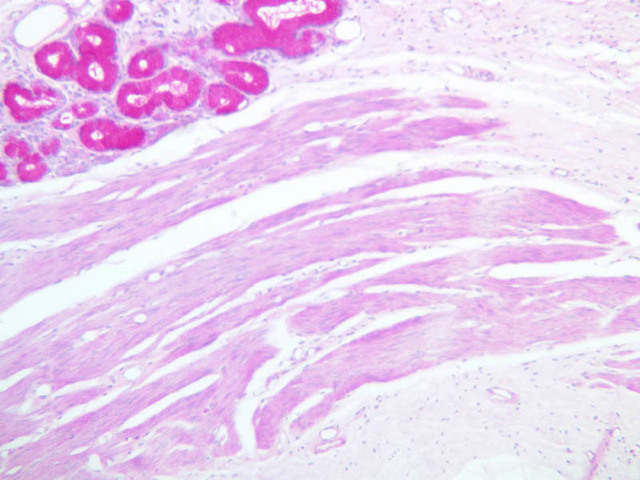
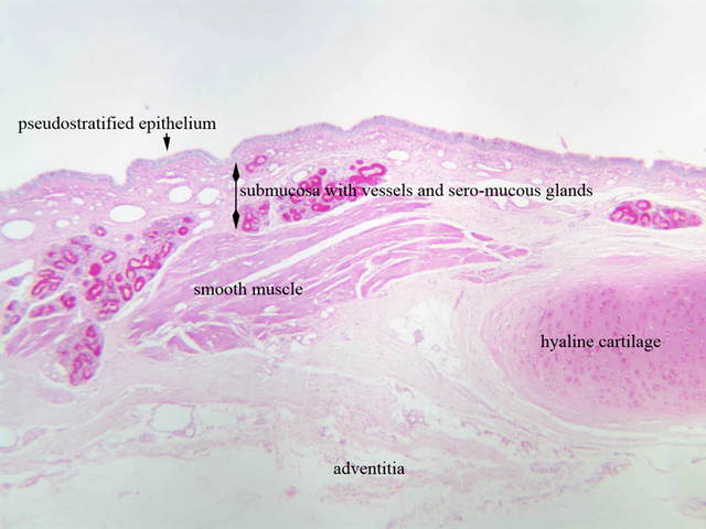
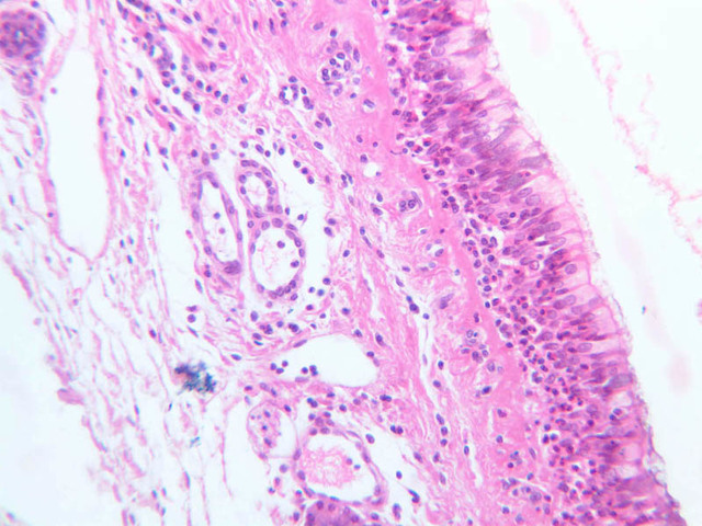
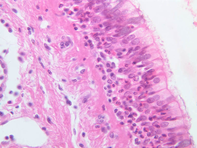
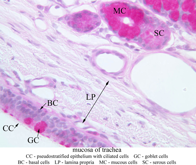
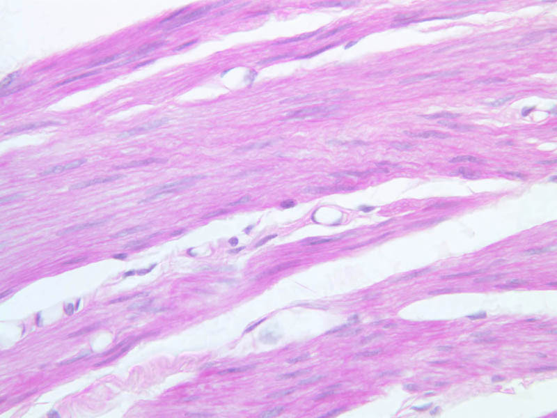
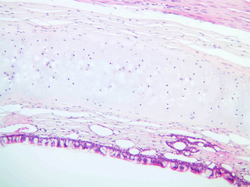
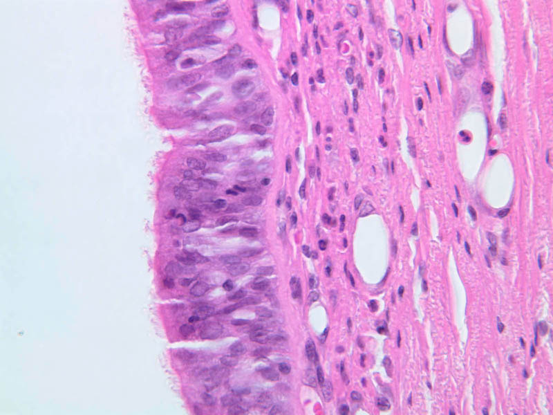
The trachea is a continuation inferiorly from the larynx (slide A-72, PAS [2.5x, 10x, 20x, 40x-labeled] [2.5x-labeled, 10x, 20x, 40x]; A-75, H&E [2.5x, 10x, 20x, 40x]; A-76, H&E [2.5x, 10x, 20x-labeled, 40x] [2.5x, 10x, 20x, 40x]). It is notable for the C-shaped rings of hyaline cartilage which keep its airway open, for the lining of mucous-coated epithelium which serves as a trap for inspired particulate matter, and for the glands beneath the epithelium which help goblet cells of the pseudostratified epithelium supply mucous. The glandular tissue may also have a role in moistening inspired air. Observe the lining epithelium with its alternating clusters of goblet and ciliated cells and the row of nuclei belonging to the basal cells. The mucosa is comprised of the epithelium and lamina propria. The lamina propria is the narrow connective layer under the epithelium. Under the lamina propria is the submucosa which contains blood vessels and seromucous glands whose ducts may be seen passing through the lamina propria. The next layer is the cartilage (or smooth muscle) layer. Hyaline cartilage rings (actually horseshoe-shaped) are covered by a dense connective tissue called the perichondrium. Note that the cartilage ring is incomplete posteriorly and that smooth muscle fibers occupy the gap. The connective tissue layer outside the cartilage rings is called the adventitia. What happens to the mucous that is produced in the trachea?Trachea Image Gallery


Trachea Table of Identifications
| Row | Structure | Abbreviation | Optimal Stain | Representative Section | Note |
|---|---|---|---|---|---|
| 1 | Ciliated Pseudostratified Epithelium | CC | PAS | |
|
| 2 | Goblet Cells | GC | PAS | |
|
| 3 | Basal Cells | BC | PAS | |
|
| 4 | Lamina Propria | LP | PAS | |
|
| 5 | Mucous Cells | MC | PAS | |
|
| 6 | Serous Cells | SC | PAS | |
|
| 7 | Submucosa | (none) | PAS | |
|
| 8 | Smooth Muscle | (none) | PAS | |
|
| 9 | Hyaline Cartilage | (none) | PAS | |
|
| 10 | Adventitia | (none) | PAS | |
|
| 11 | Seromucous Glands | (none) | H&E | |
|
| 12 | Perichondrium | (none) | H&E | |
Top of page
Lung
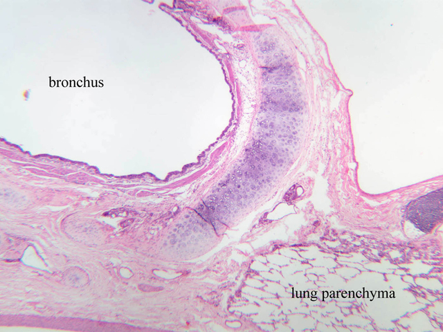
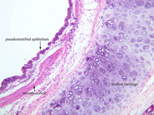
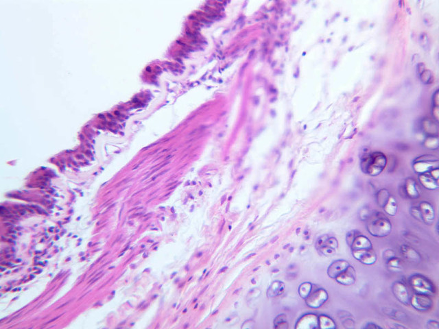
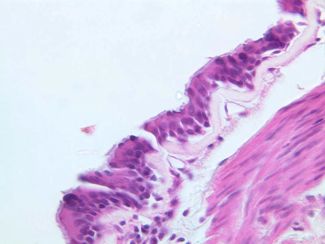
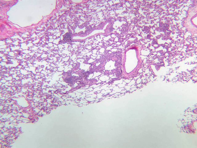
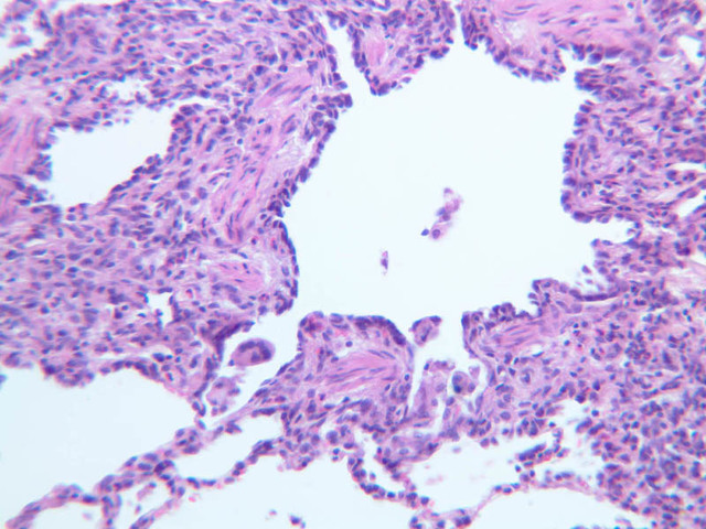
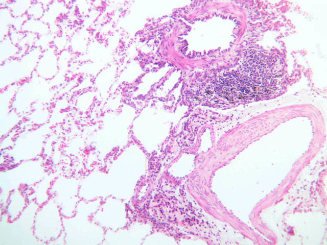
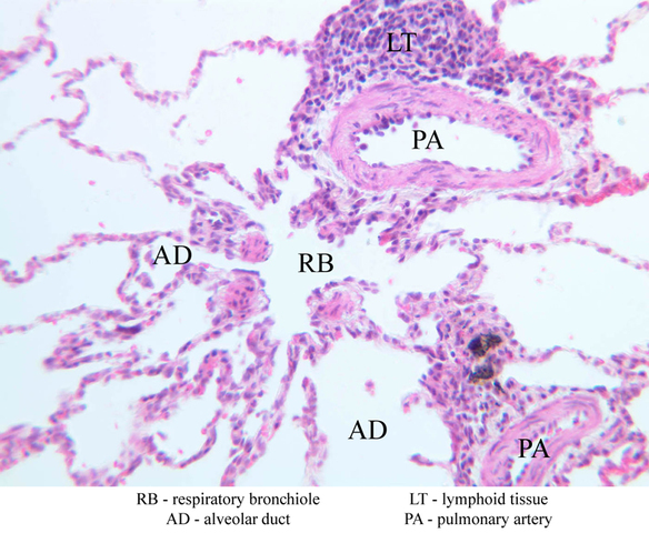
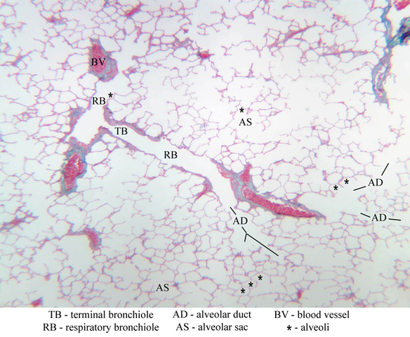
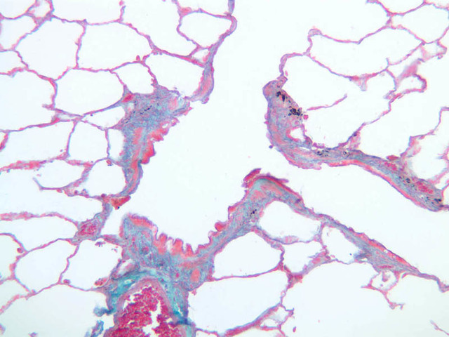
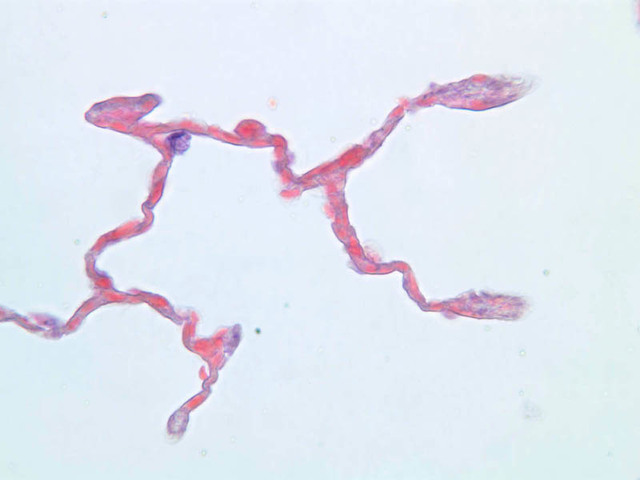
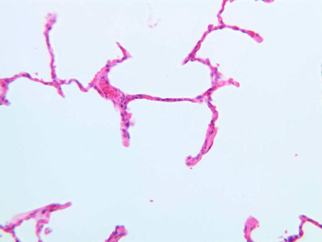
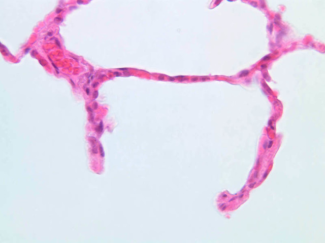
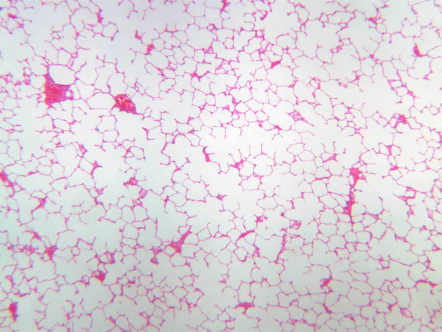
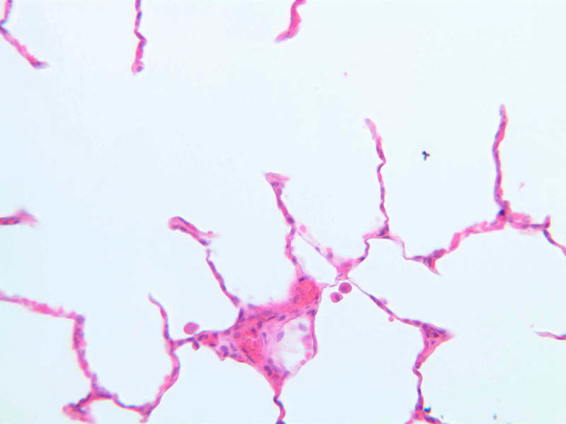
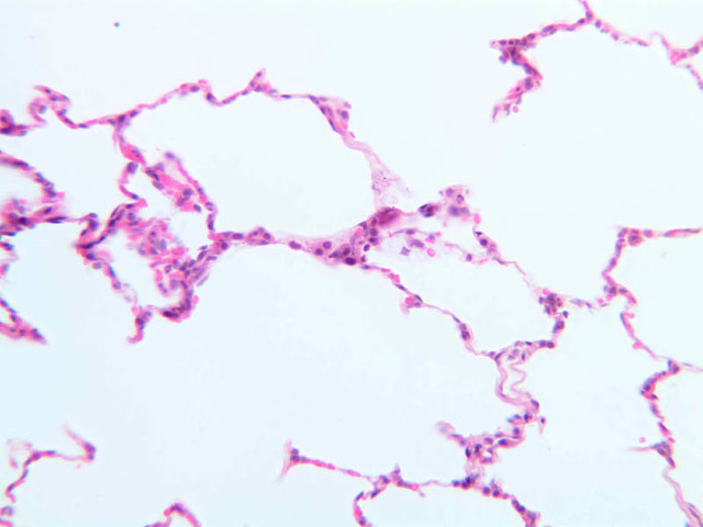
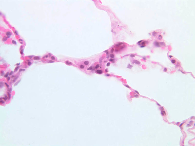
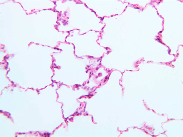
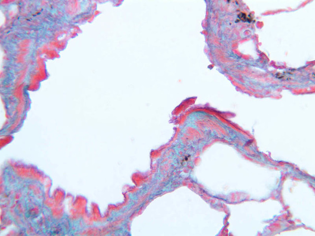
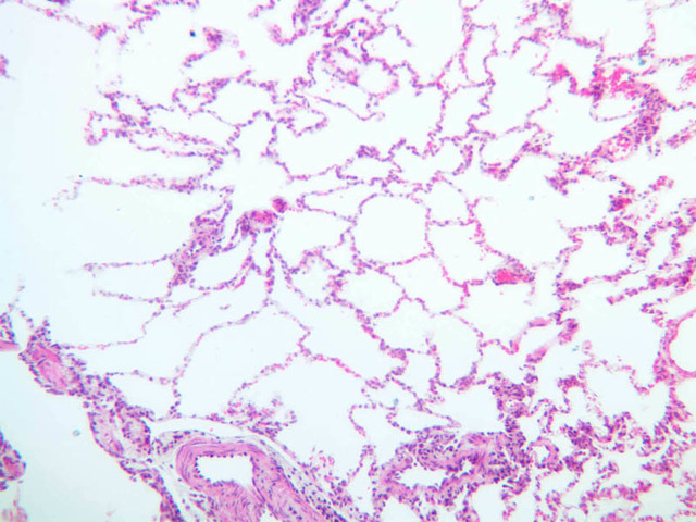
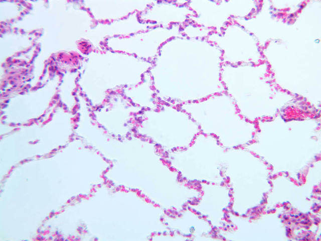
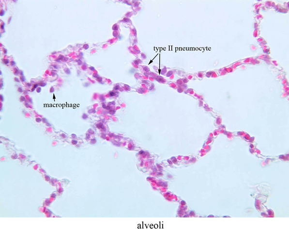
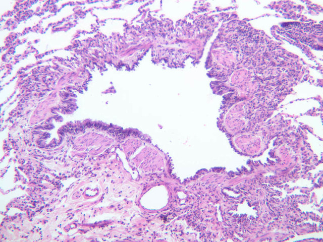
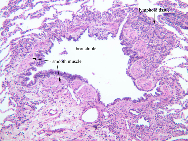
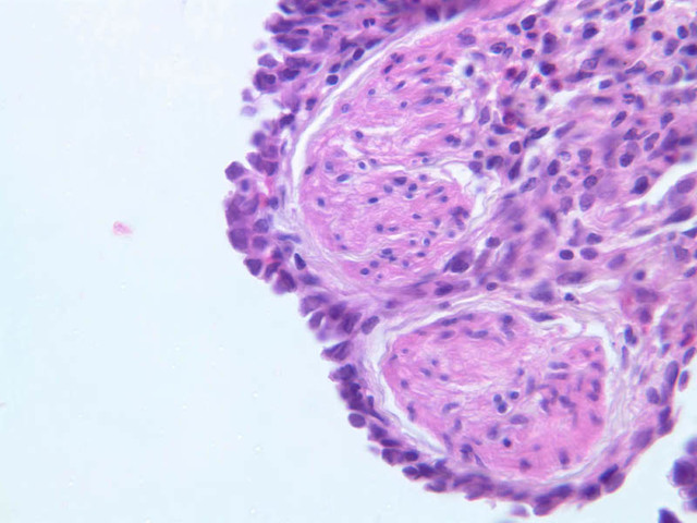
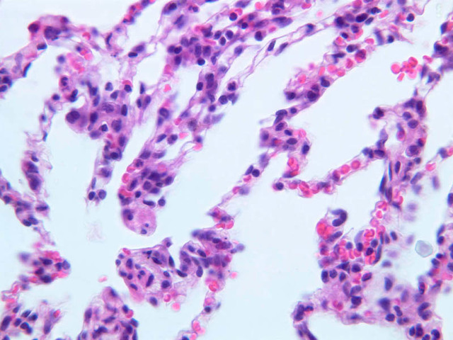
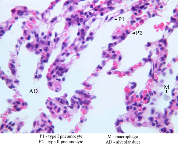
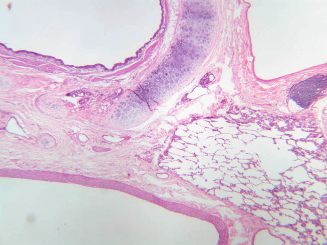
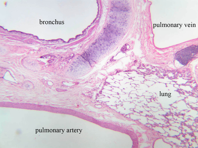
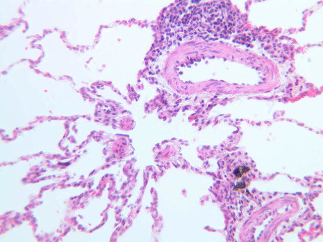
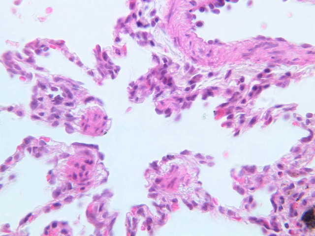
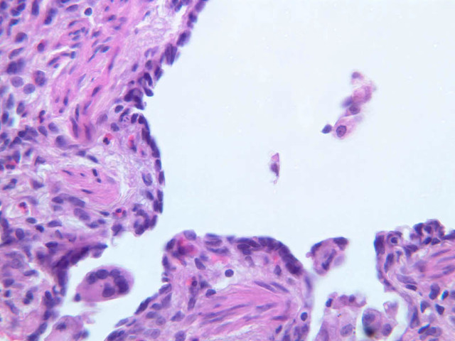
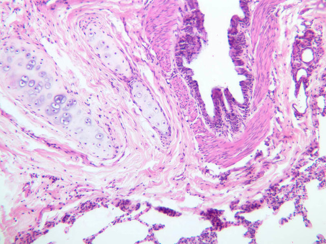
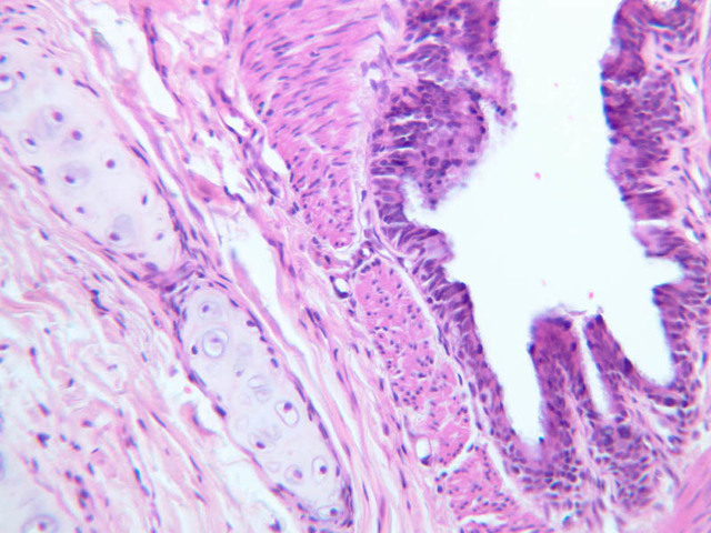
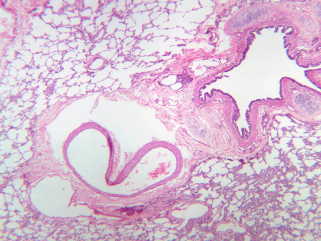
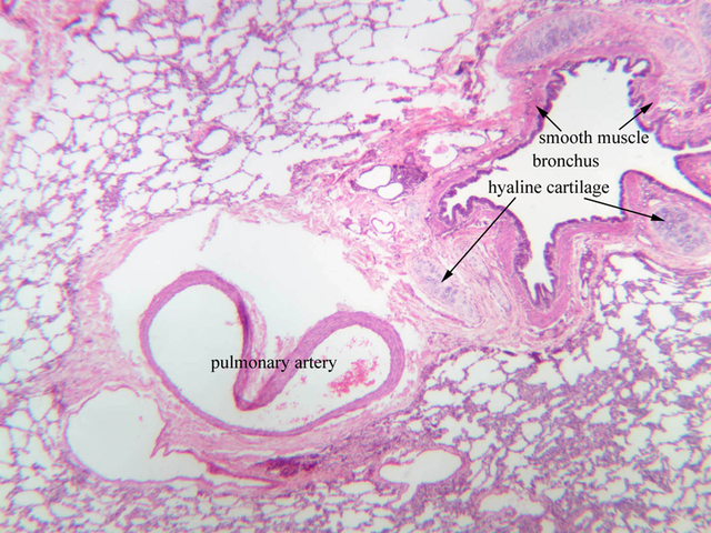
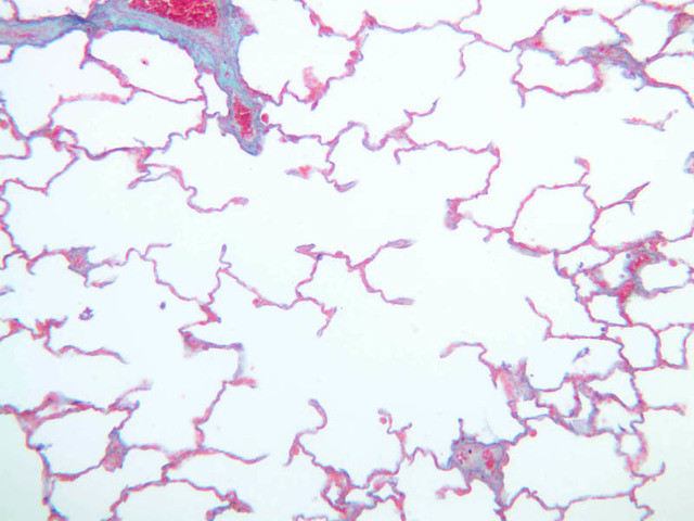
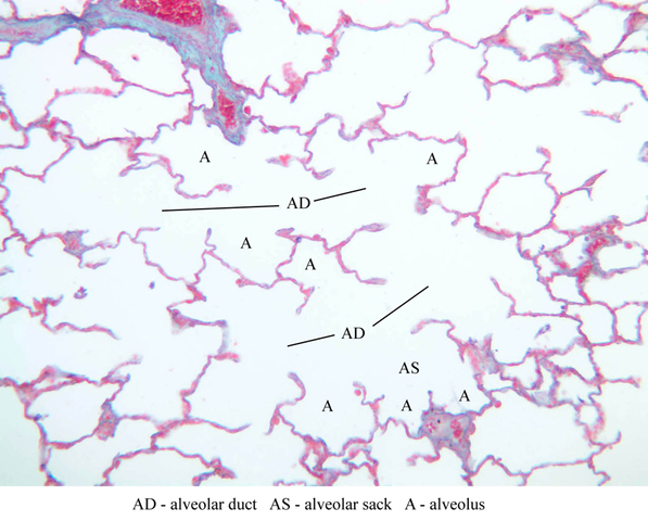
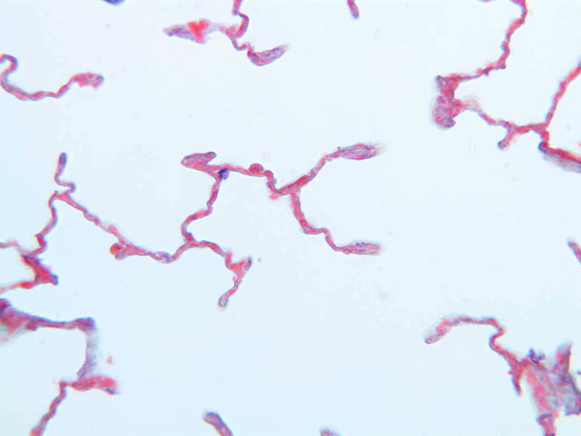
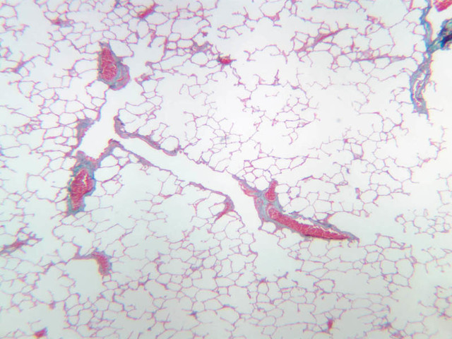
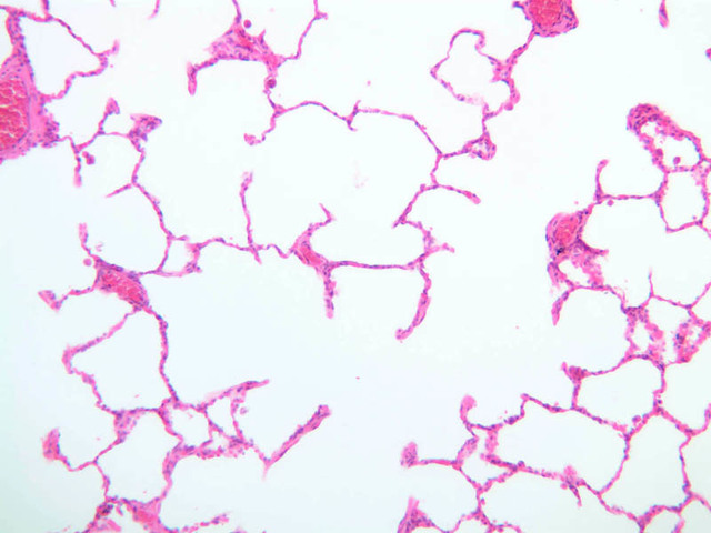
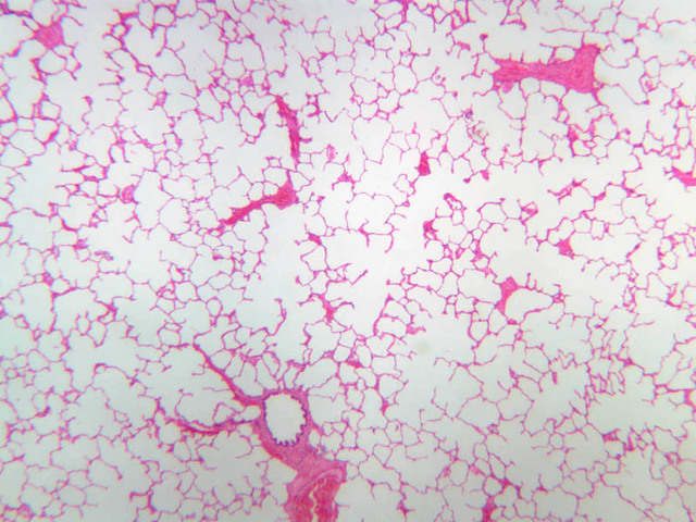
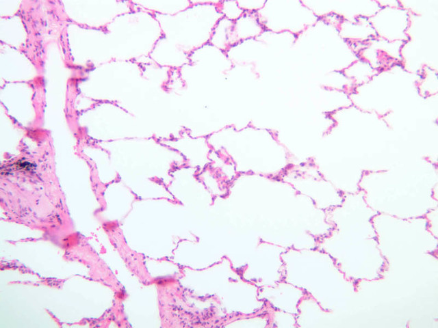
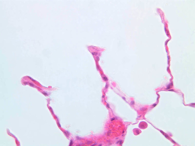
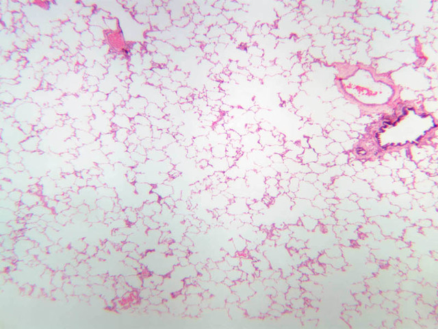
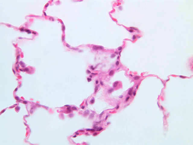
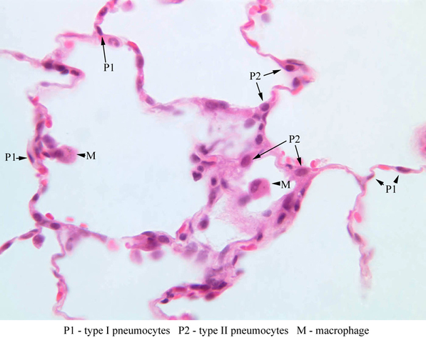
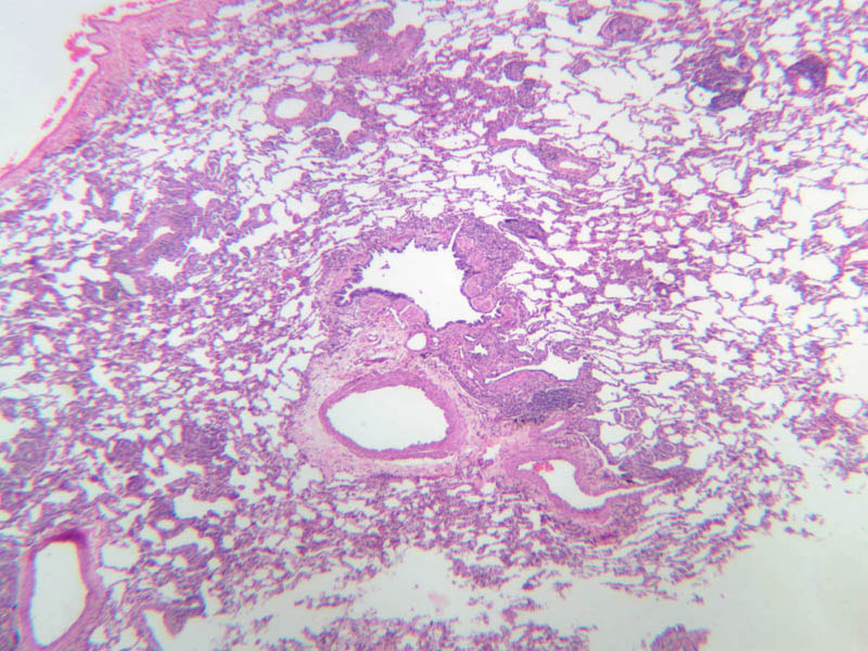
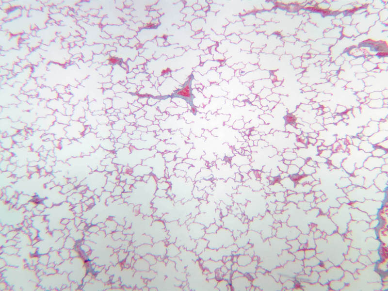
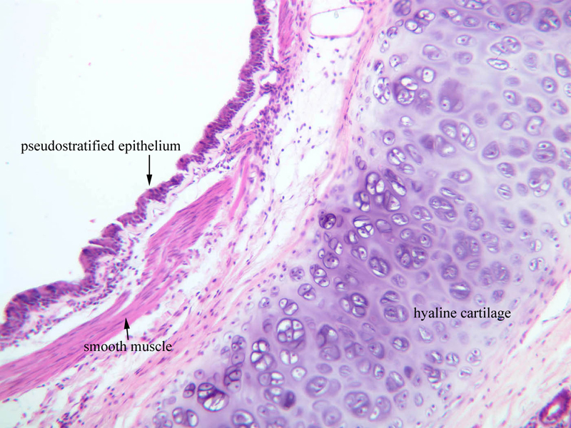
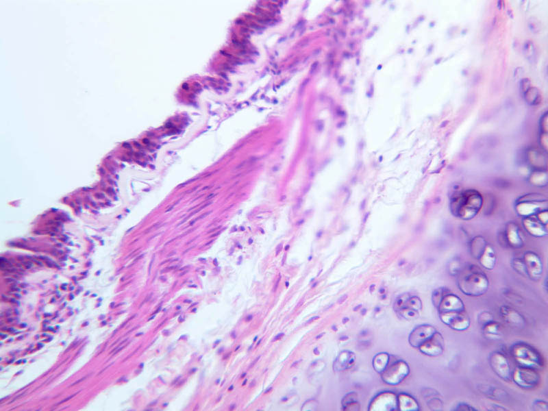
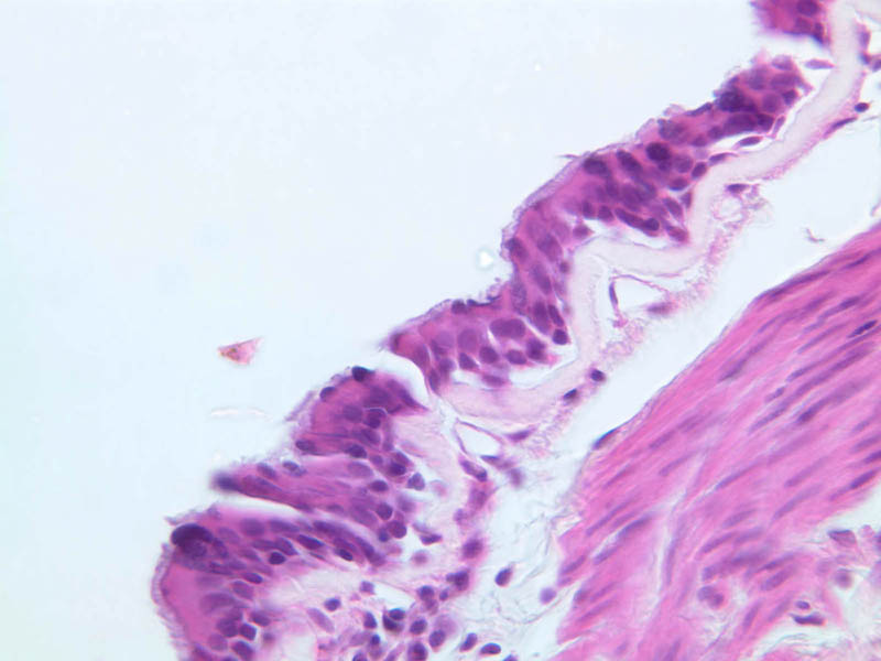
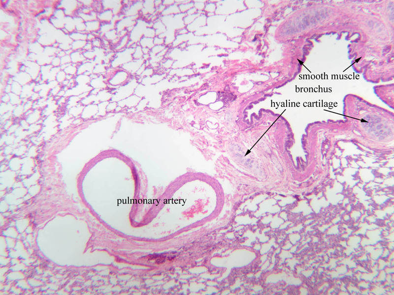
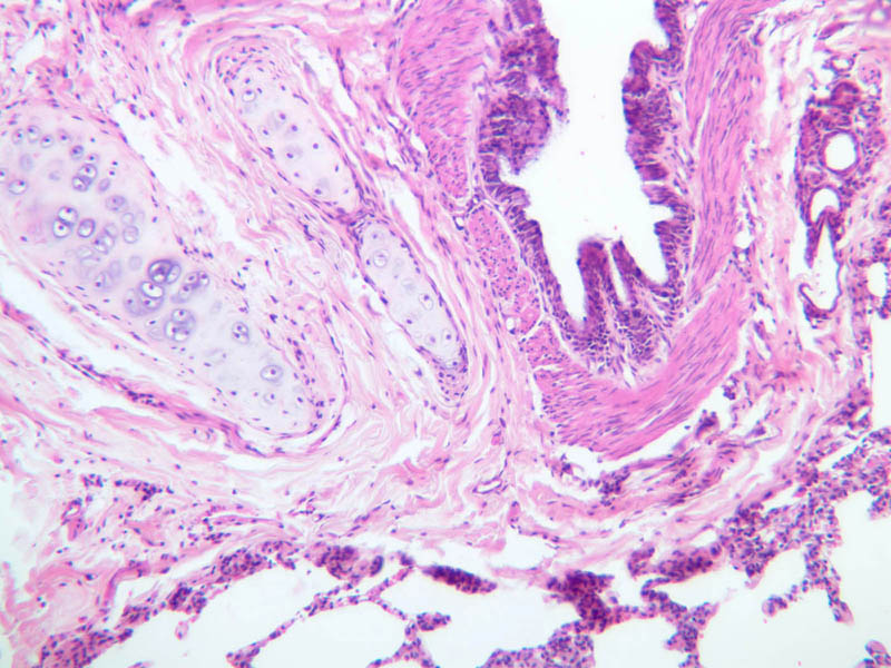
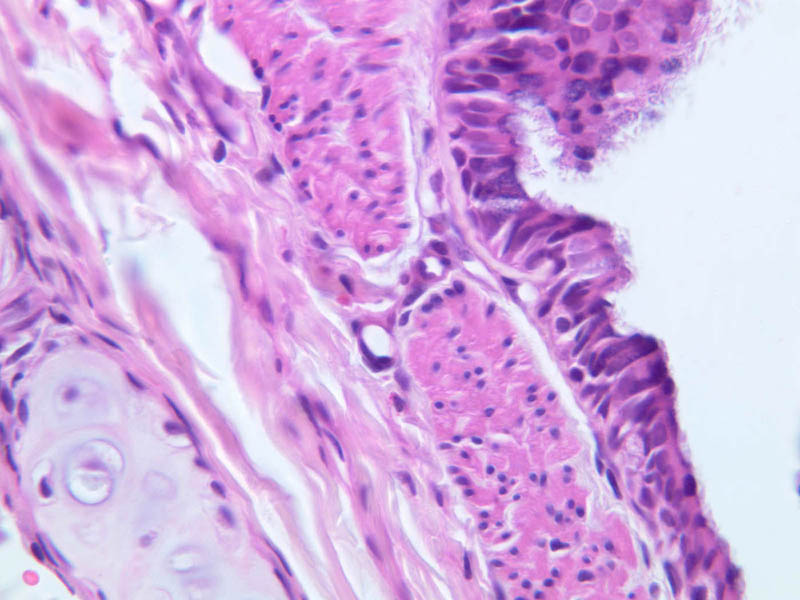
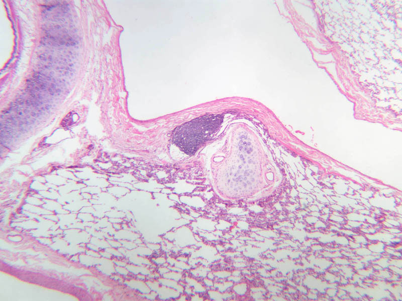
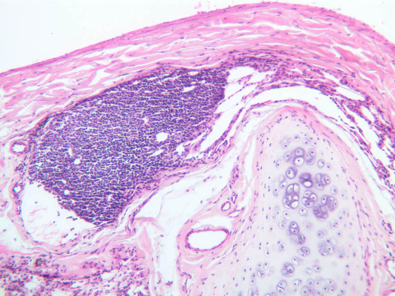
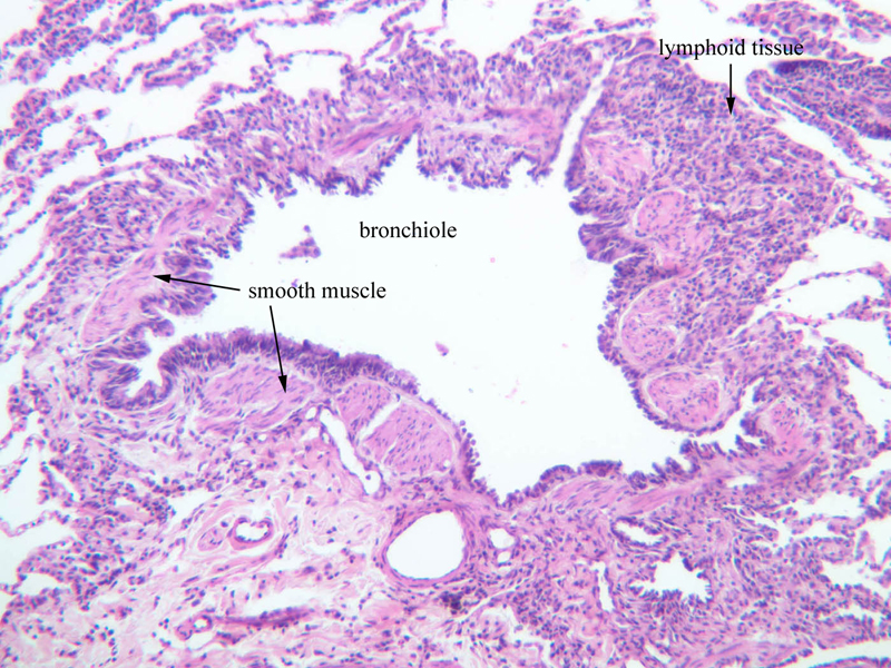
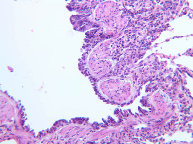
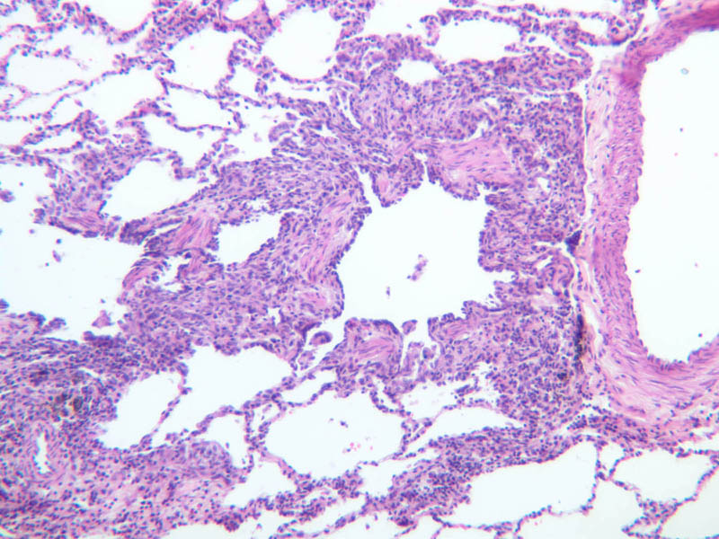
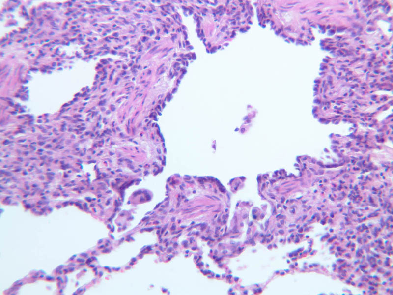
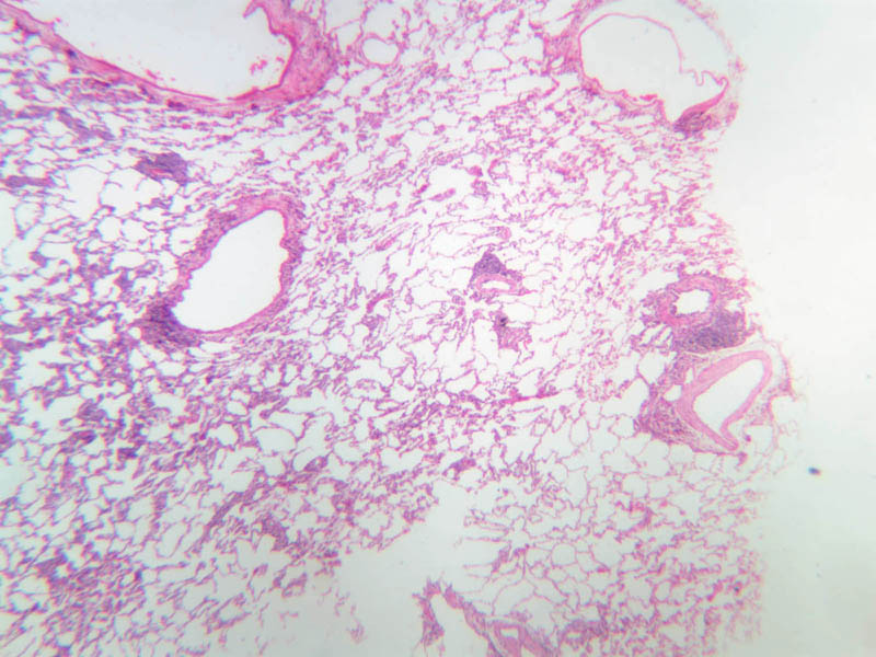
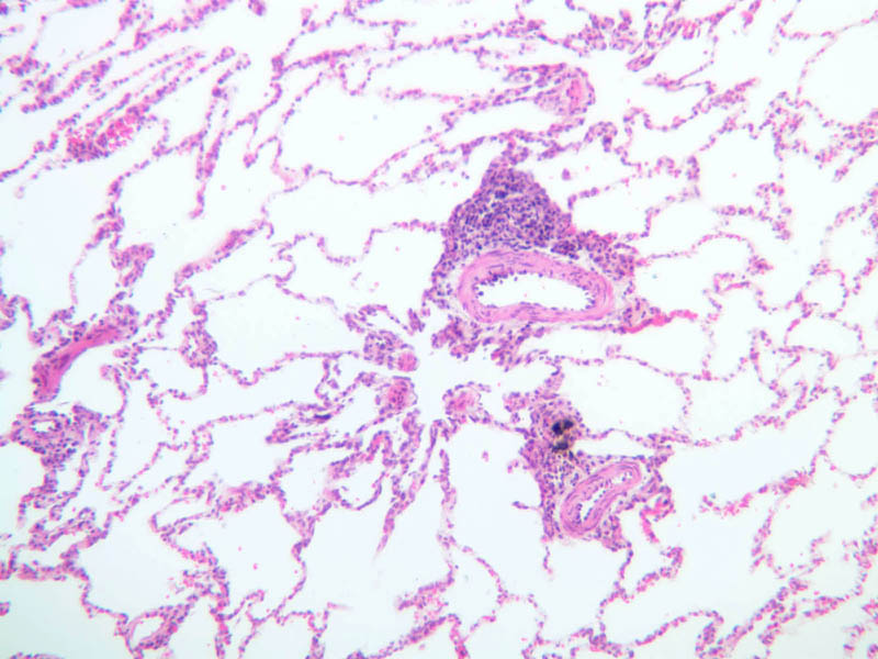
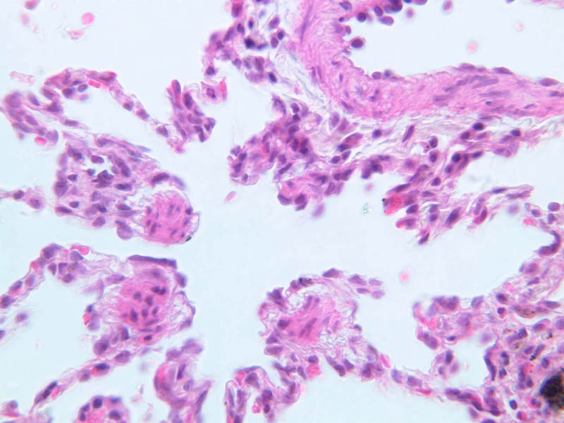
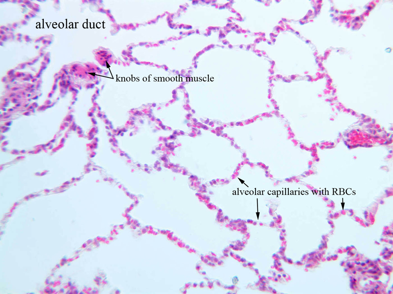
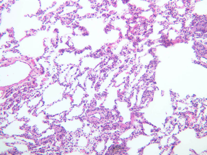
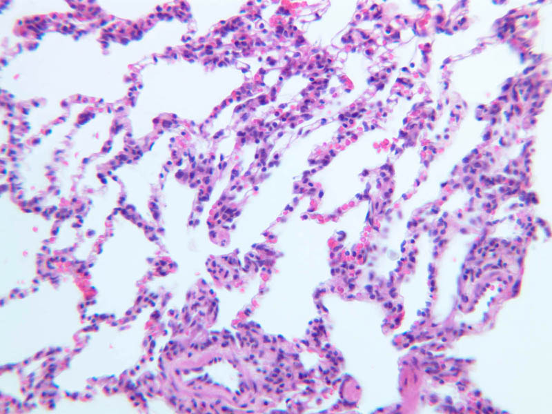
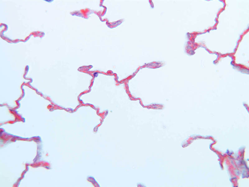
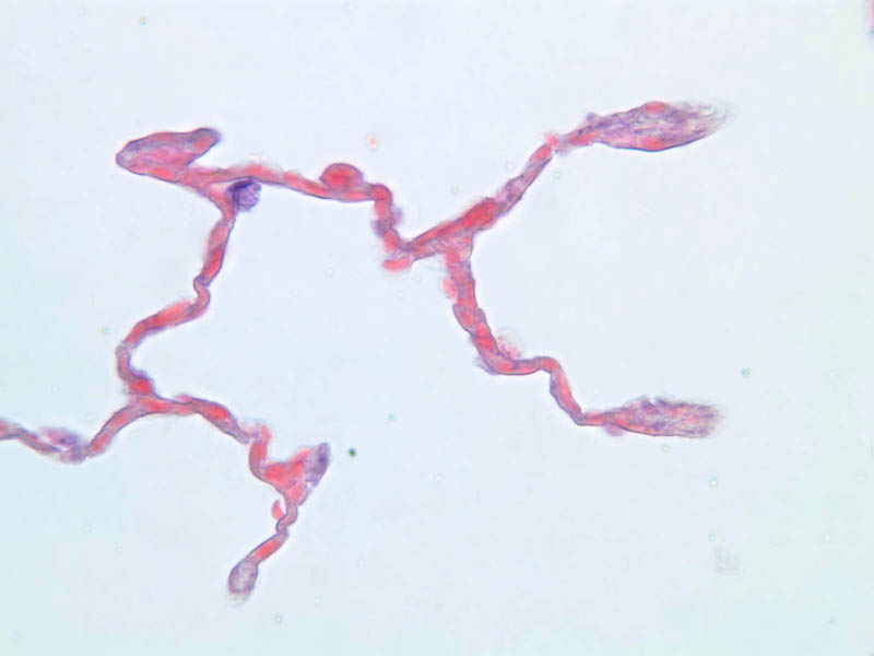
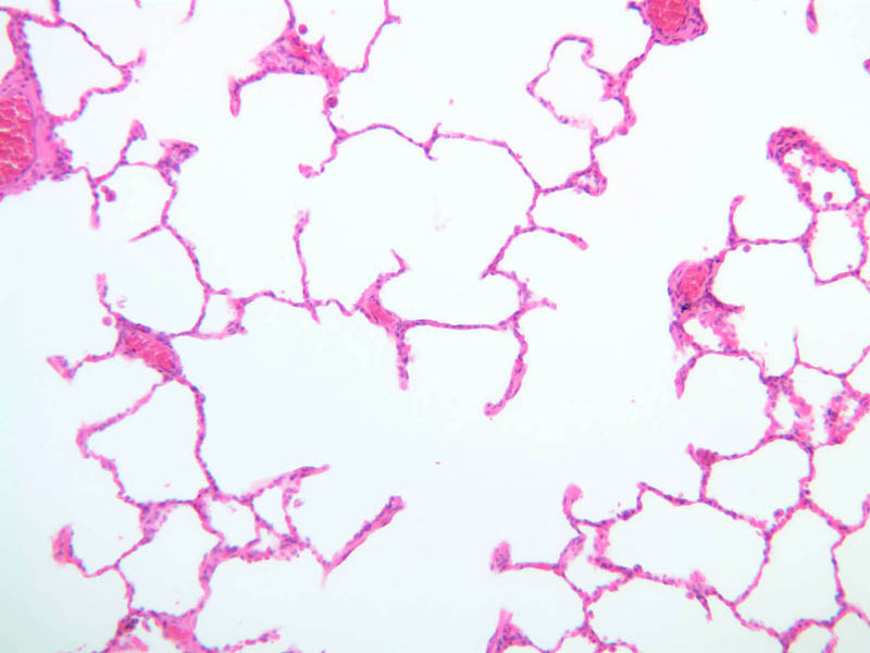
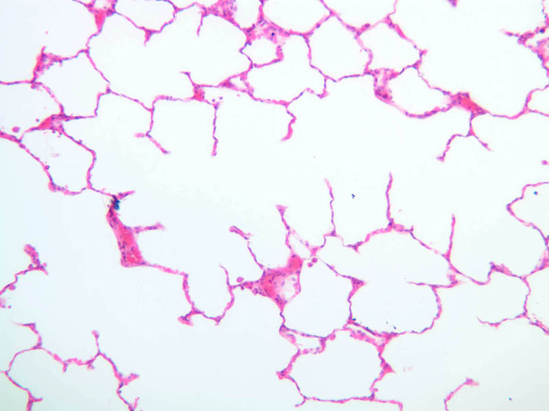
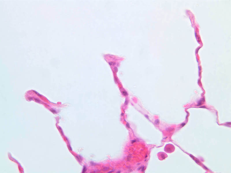
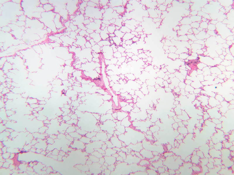
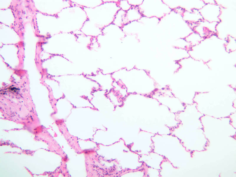
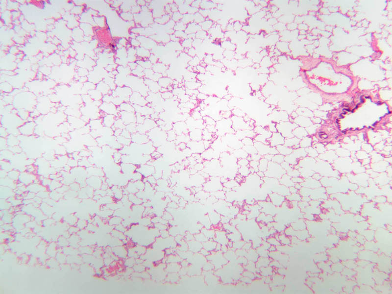
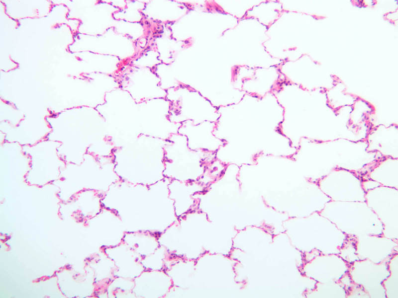
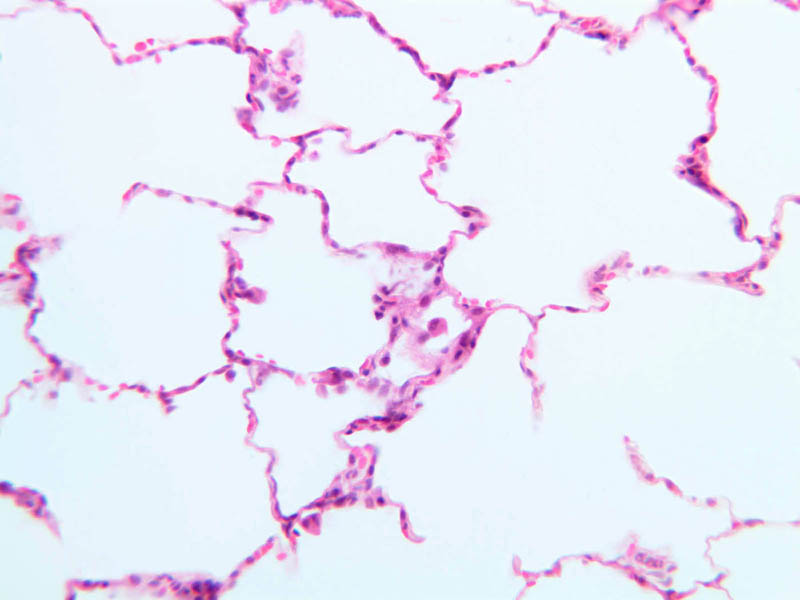
Orientation: Before attempting to examine the class set specimens, first review and study lung morphology in your text and atlas. Understand the morphology and relative size of 1) bronchi; 2) bronchioles; 3) respiratory bronchioles; 4) alveolar ducts; 5) alveolar sacs; and 6) alveoli. In these micrographs, trace the passage of a molecule of oxygen from the bronchi to blood. Doing this beforehand will greatly facilitate examination of the specimens. Examine slides A-74 (H&E; lung and bronchi), A-77 (AF; lung), and A-78 (H&E; lung); refer to the micrographs indicated above. The trachea enters the thorax and bifurcates, giving rise to two primary (extrapulmonary) bronchi. Each primary bronchus enters a lung and branches repeatedly to form smaller bronchi (A-74 [2.5x-labeled, 10x-labeled, 20x, 40x] [2.5x-labeled, 10x, 20x, 40x]). Primary bronchi resemble the trachea, having cartilages to support the tubes and keep them open and pseudostratified epithelium as an epithelial lining. However, the ring-shaped cartilages of the trachea and primary bronchi give way to irregular and discontinuous plates of cartilages in the smaller bronchi. Also, as the bronchi enter the lungs, strands of smooth muscle appear, oriented circularly around the airway within the cartilage shell, but forming an incomplete ring. As bronchi divide and form smaller tubes, the cartilage plates diminish in number and the relative amount of muscle around the tubes increases. In part, because of the presence of muscle, the mucosa of the bronchi tends to have a corrugated appearance in cross-section. Besides the increase in muscular tissue, lymphoid tissue occurs frequently along the bronchial tubes (A-74 [2.5x, 10x, 20x, 40x]). Its importance with regard to pathogens brought in with inspired air and trapped by the mucous coat of the epithelium should be obvious. The epithelial lining itself gradually becomes lessened in height as the airways decrease in diameter. Ciliated cells and goblet cells continue, however, as important constituents of the epithelium. As the air passages bifurcate, they become smaller and lose their cartilage plates. Several orders of smaller and smaller bronchioles are thus formed (A-74 [2.5x, 10x-labeled, 20x, 40x] [2.5x, 10x, 20x, 40x] [10x]). In these tubes, muscle and elastic tissue form an incomplete feltwork of tissue around the luminal epithelium. Multicellular glands are usually not present in bronchioles, the goblet cells being sufficient to provide the mucous coat, but lymphoid tissue continues to occur in patches along these small airways. The height of epithelial cells decreases until only a layer of cuboidal cells remains as the lining of the terminal bronchioles. The elastic and muscular coats shorten and decrease the caliber of bronchioles during expiration. Also, acute spasms of the musculature, such as may occur in allergic and anaphylactic reactions, can bring about suffocation. The final branches of the respiratory tree occur at the level of the terminal bronchiole. These branches give rise to the primary (or functional) lobule of the lung. It consists of a respiratory bronchiole (A-74 [2.5x, 10x, 20x-labeled, 40x]; A-77 [2.5xlabeled, 10x, 20x, 40x]), whose wall is thinned and evaginated from place to place to form solitary respiratory alveoli, and its further subdivisions of alveolar ducts, atria, alveolar sacs and alveoli, together with attendant arteries and capillaries. Look for a longitudinal section through a primary lobule and identify these various subdivisions (A- 74 [10x, 20x-labeled, 40x-labeled] [10x, 20x, 40x-labeled]; A-77 [2.5x, 10x-labeled, 20x, 40x]; A-78 [2.5x, 10x, 20x, 40x] [2.5x, 10x, 20x, 40x]). The paths of alveolar ducts can be followed by observing knobs of smooth muscle tissue covered by squamous epithelium which appear to project into the airway. Alveolar sacs and alveoli open along the path of the alveolar duct and make its wall appear to be discontinuous. Study the architecture of the alveoli. Look for alveolar macrophage cells and type II cells. The macrophages (also known as dust cells) may contain prominent granules of ingested material. It is very difficult to distinguish individual cells in the alveolar wall with the light microscope. As in other parts of the respiratory system, the epithelium contains two predominant cell types. The type I pneumocyte is a large and highly attenuated cell that covers most of the alveolar surface. The type II pneumocyte (great alveolar cell; granular pneumocyte; septal cell) is a secretory cell that is responsible for producing pulmonary surfactant (A-78 [2.5x, 10x, 20x, 40x] [2.5x, 10x, 20x, 40x-labeled]). The type II cell also functions as a stem cell after injury and divides to form new epithelial cells that differentiate into type I cells and re-establish the epithelial surface. Study the alveolar walls and note that they are filled with capillaries. The capillaries are surrounded by delicate elastic tissue. Though contraction of the major air ducts during expiration is a chief role of elastic tissue in the lung, the presence of elastic fibers in the alveolar walls suggests these are stretched during inspiration and contract during expiration. Review the relationship of blood vessels to the conducting passages of the lung. A branch of the pulmonary artery enters the lung at its hilus with the primary bronchus. Subdivisions of the artery then follow and are loosely attached to bronchial and bronchiolar airways. Pulmonary arterioles terminate in the extremely dense capillary bed of the lungs at the level of alveolar ducts. Look for examples of these vessels (A-74 [2.5x-labeled, 10x, 20x, 40x]). From the alveolar capillary bed, the capillaries join to form venules and veins that run as solitary vessels through the connective tissue between pulmonary lobules back towards the hilus. Find examples of these veins. The barrier that lies between air and blood at the level of the alveolus is clearly demonstrated in an electron micrograph of the alveolar wall. The lining of both the alveolus and the capillary is complete and air traverses these cellular layers and their basement laminae (often fused) in the respiratory exchange. How many cells must a molecule of oxygen cross from the alveolar space to the capillary lumen? How many unit membranes would be traversed in this journey?Lung Image Gallery


Lung Table of Identifications
| Row | Structure | Abbreviation | Optimal Stain | Representative Section | Note |
|---|---|---|---|---|---|
| 1 | Bronchus | (none) | H&E | |
|
| 2 | Lung Parenchyma | (none) | H&E | |
|
| 3 | Pseudostratified Epithelium | (none) | H&E | |
|
| 4 | Smooth Muscle | (none) | H&E | |
|
| 5 | Hyaline Cartilage | (none) | H&E | |
|
| 6 | Pulmonary Artery | (none) | H&E | |
|
| 7 | Bronchiole | (none) | H&E | |
|
| 8 | Lymphoid Tissue | (none) | H&E | |
|
| 9 | Respiratory Bronchiole | RB | H&E | |
|
| 10 | Alveolar Duct | AD | H&E | |
|
| 11 | Terminal Bronchiole | TB | AF | |
|
| 12 | Alveolar Sac | AS | AF | |
|
| 13 | Blood Vessel | BV | AF | |
|
| 14 | Alveoli | (*), A | AF | |
|
| 15 | Knobs of Smooth Muscle | (none) | H&E | |
|
| 16 | Alveolar Capillaries (with RBCs) | (none) | H&E | |
|
| 17 | Macrophage | (none), M | H&E | |
|
| 18 | Type II Pneumocyte | P2 | H&E | |
|
| 19 | Type I Pneumocyte | P1 | H&E | |
|
| 20 | Pulmonary Vein | (none) | H&E | |
|
| 21 | Lung | (none) | H&E | |
Top of page
Stages of Lung Development
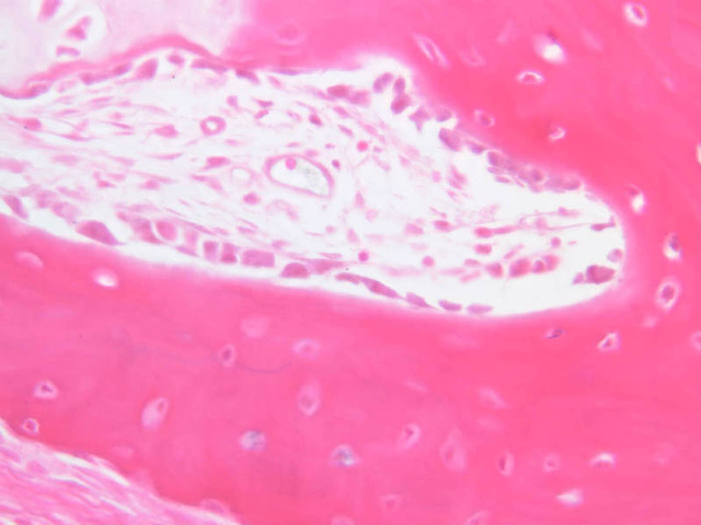
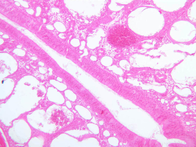
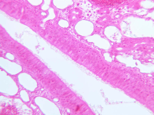
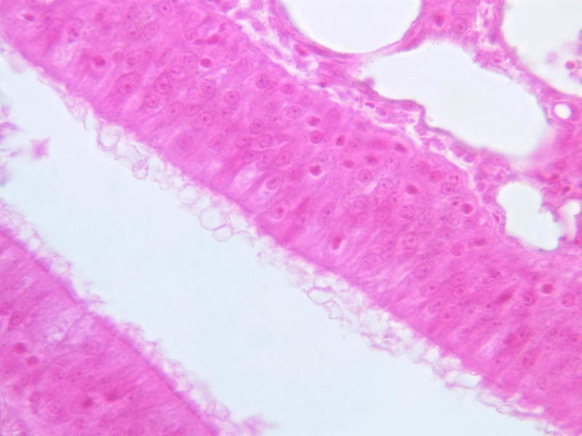
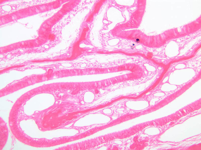
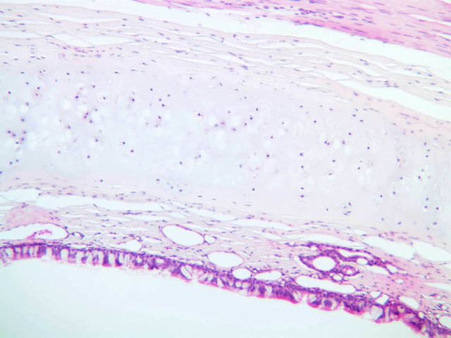
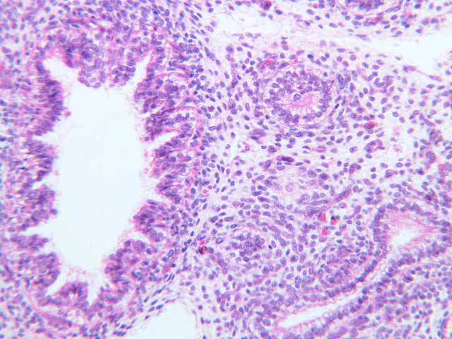
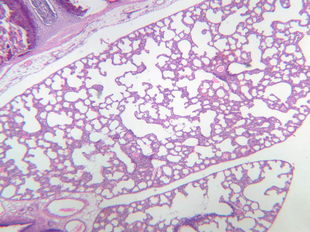
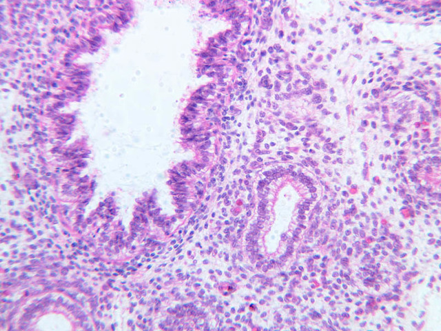
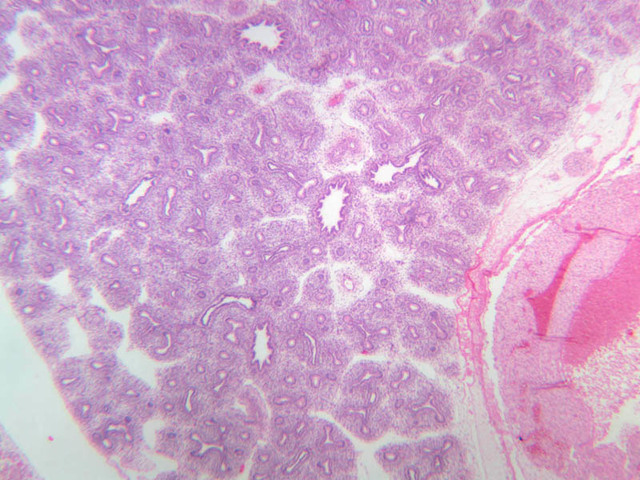
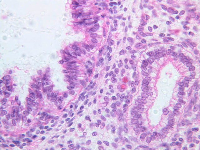
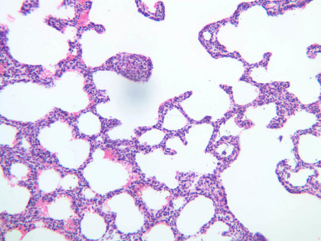
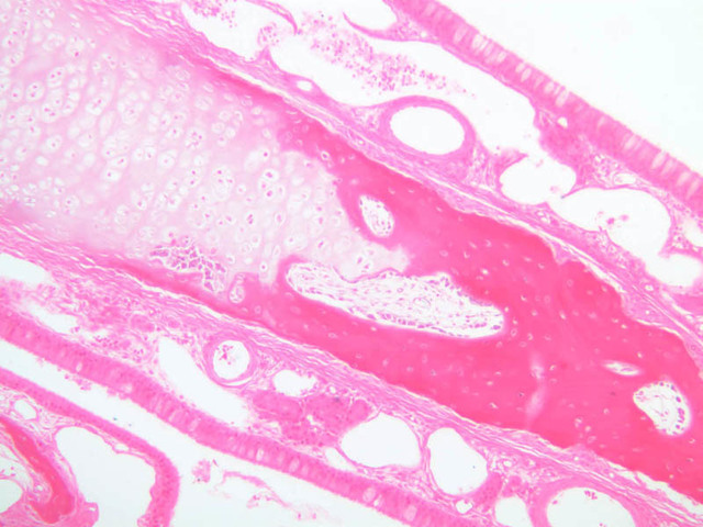
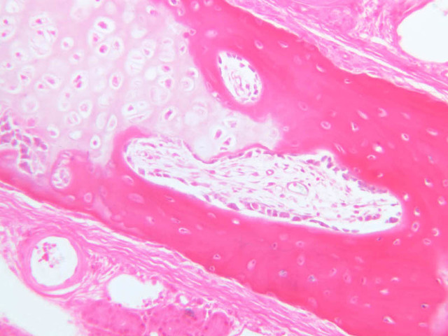
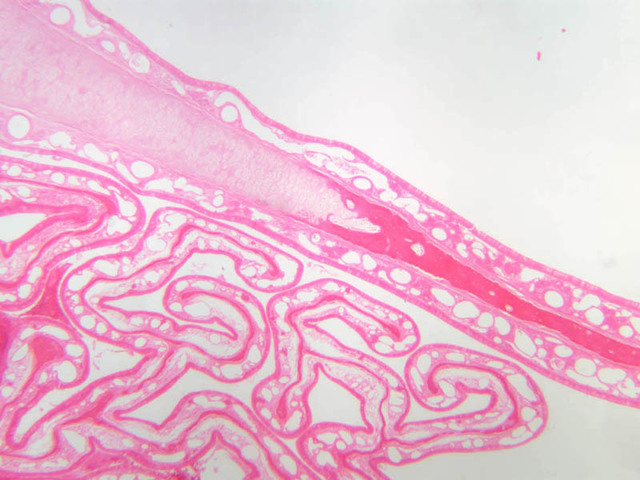
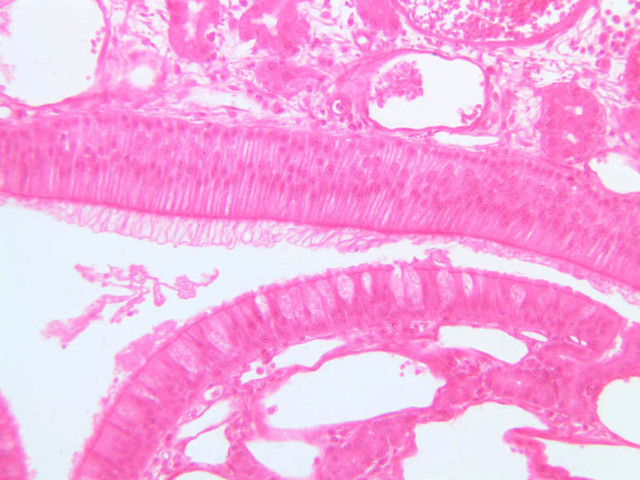
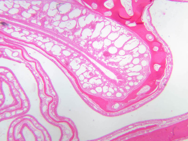
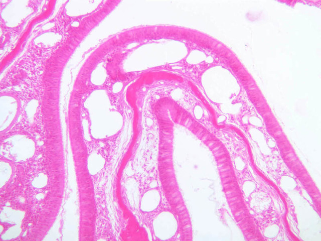
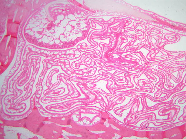
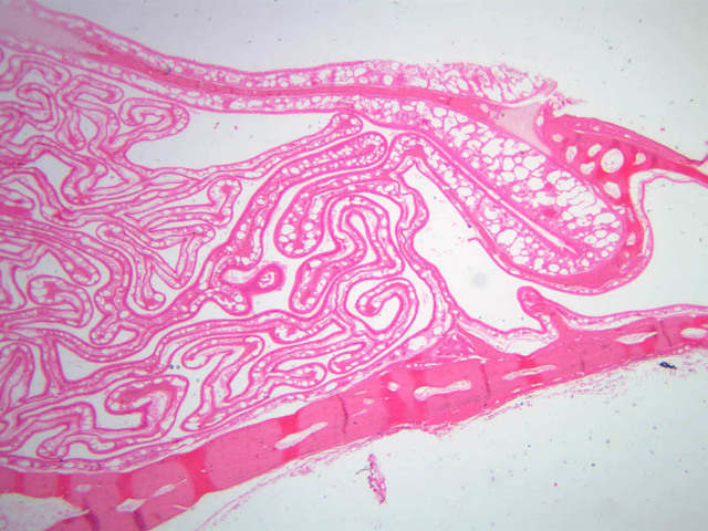
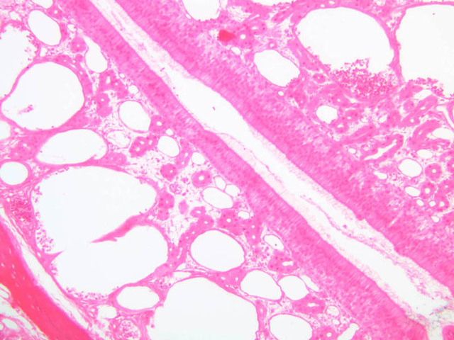
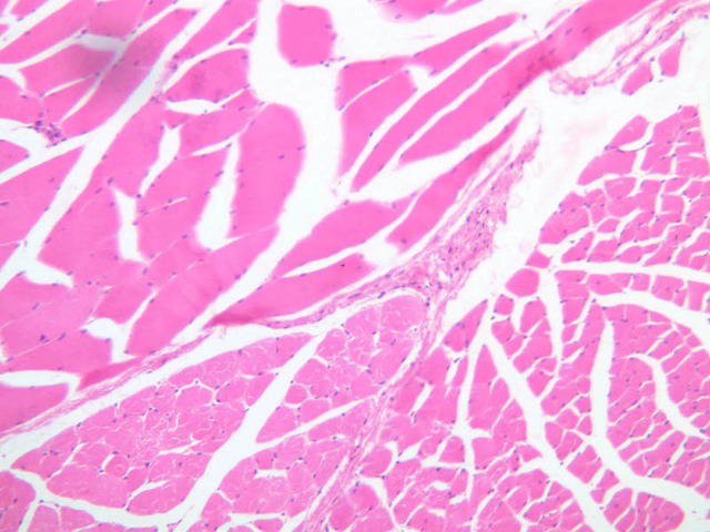
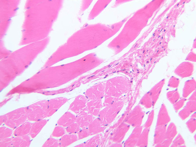
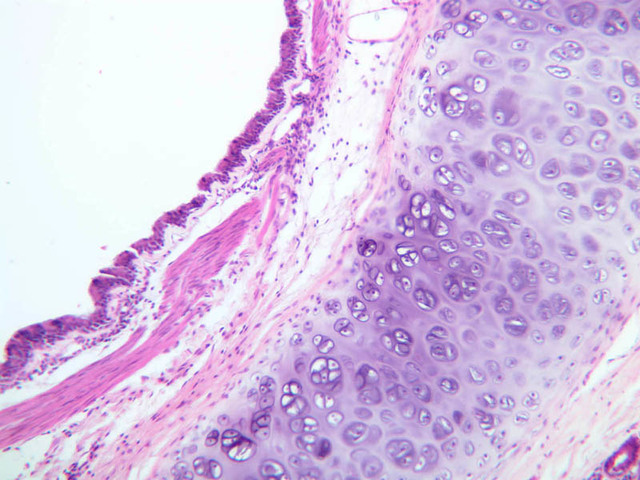
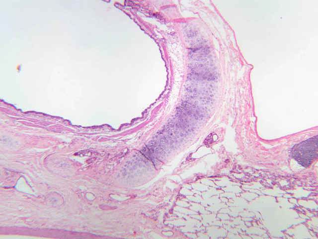
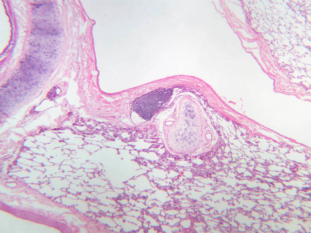
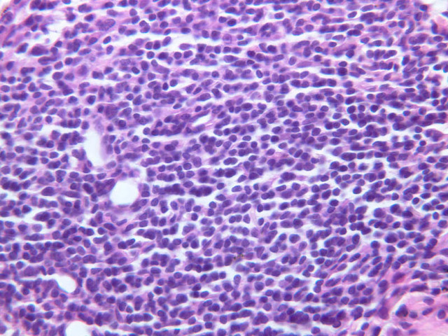
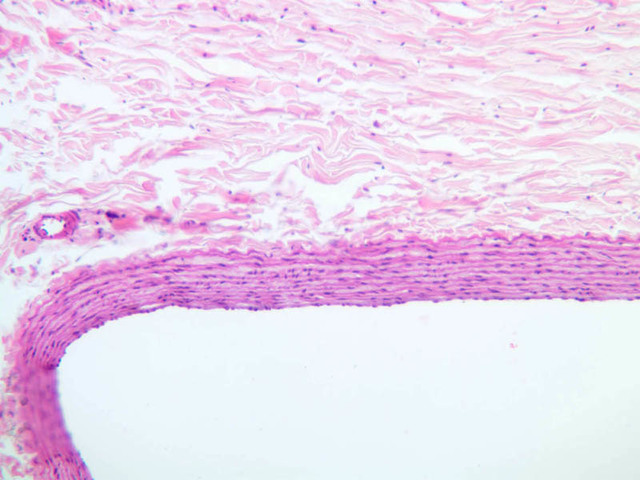
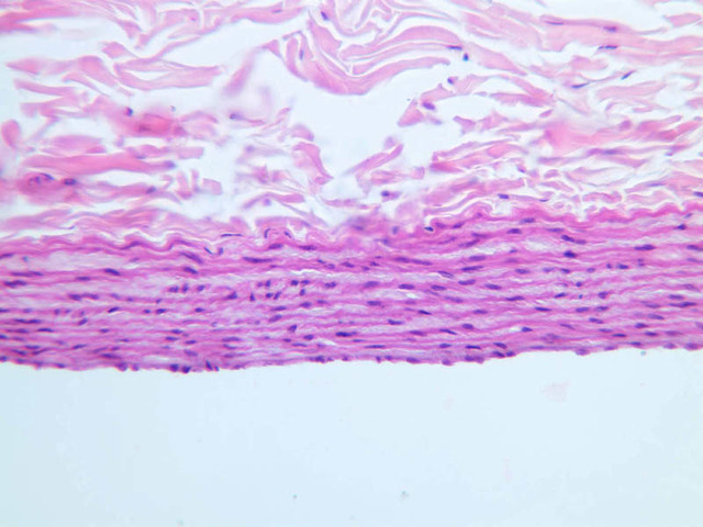
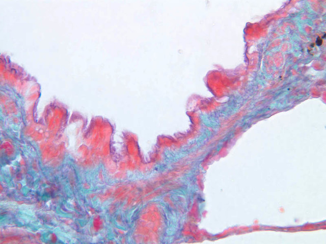
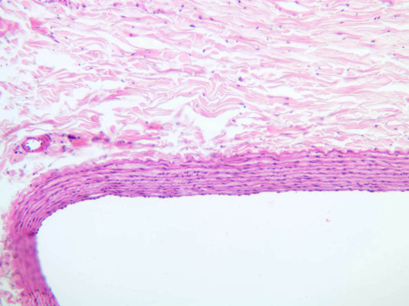
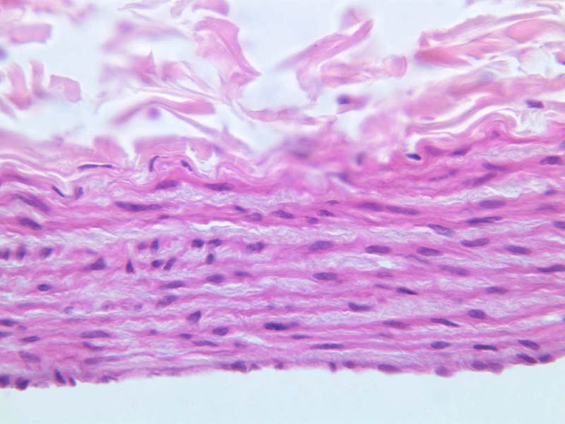
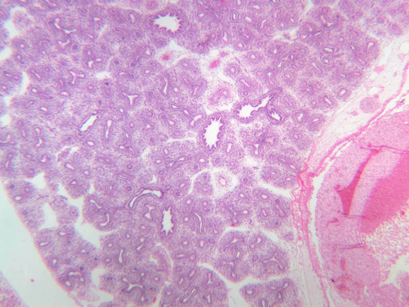
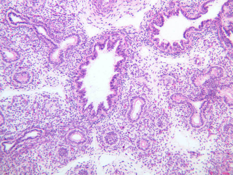
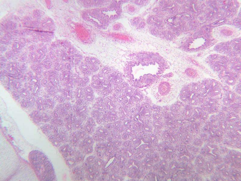
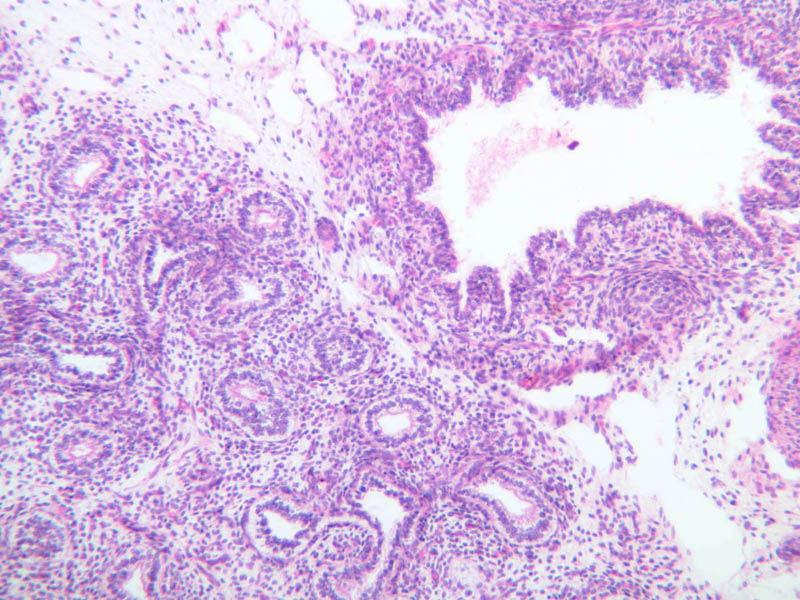
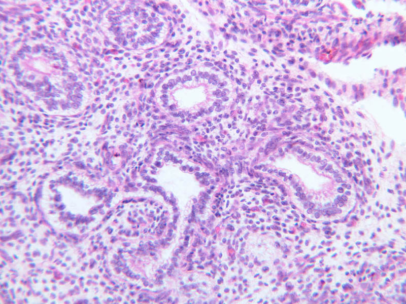
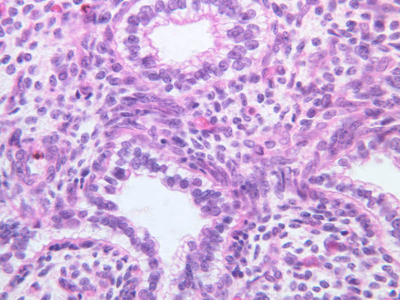
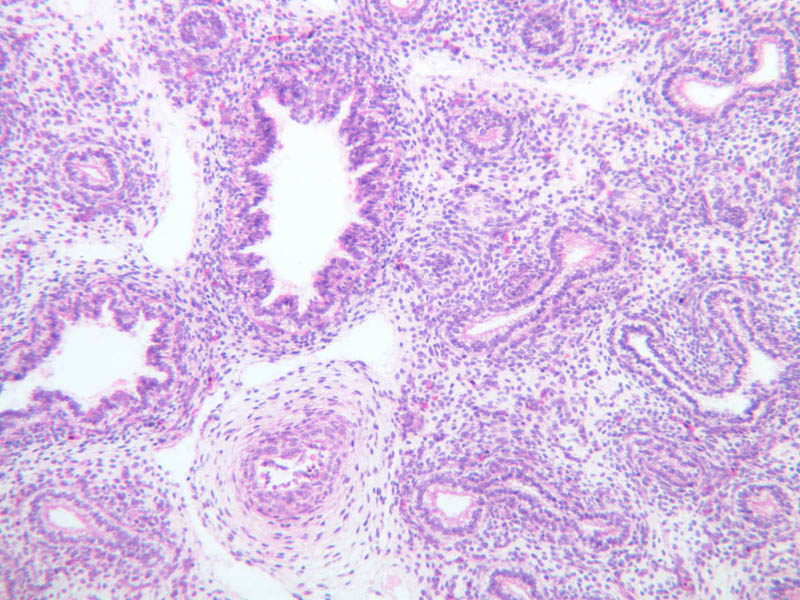
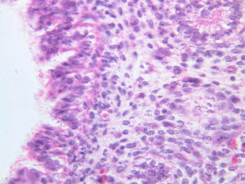
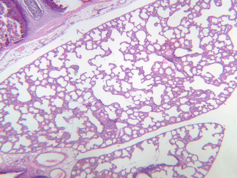
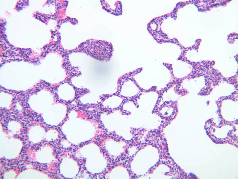
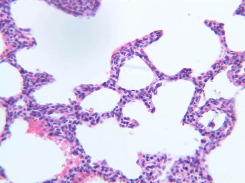
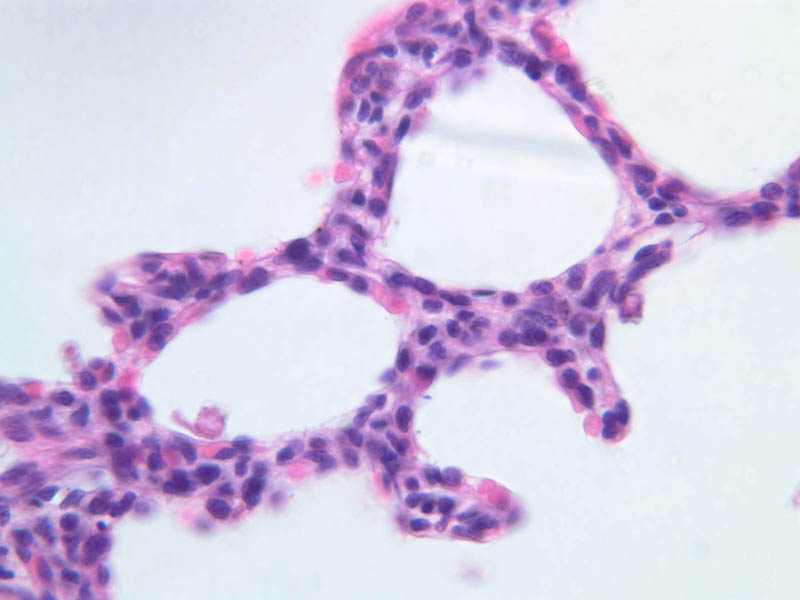
There are four stages of lung development; three are prenatal and the final is postnatal. Review your notes after the lecture on lung development and study the limited class set material. I. Pseudoglandular (weeks 5 to 17) - slide A-13 ([2.5x, 10x, 20x, 40x] [2.5x, 10x, 20x, 40x] [10x, 20x, 40x]). At this stage of development, future air spaces are lined by columnar or cuboidal epithelium and resemble glands. However, the major structural features of the lung are all present and the relationship between airways and branches of pulmonary artery and vein are readily apparent. II. Canalicular (weeks 16 to 25). During this stage airway lumina increase in size, differentiation of the epithelium begins, there is a significant increase in vascularization and the walls of some primitive alveoli or terminal sacs thin out enough to permit gas exchange if premature birth occurs. III. Terminal sac (weeks 24 through birth) - slide A-14 ([2.5x, 10x, 20x, 40x]). In the terminal sac stage alveolar development continues. Alveolar epithelial differentiation progresses and alveolar walls are thin enough to permit gas exchange. In the later part of this stage, surfactant production begins in preparation for birth. IV. Alveolar (late fetal to about 8 years). In the alveolar stage, there is continued thinning of the alveolar system and further septation occurs resulting in the formation of many alveoli from each terminal sac.Lung Development Image Gallery


Top of page
Chapter Nine Review
Review of Slides


Review of Identifications
| Row | Structure | Abbreviation | Optimal Stain | Representative Section | Note |
|---|---|---|---|---|---|
| 1 | Olfactory Epithelium | (none) | H&E | |
|
| 2 | Respiratory Epithelium | (none) | H&E | |
|
| 3 | Turbinate Bone | (none) | H&E | |
|
| 4 | Network of Veins | (none) | H&E | |
|
| 5 | Nonmotile Cell | (none) | H&E | |
|
| 6 | Supporting Cell | (none) | H&E | |
|
| 7 | Olfactory Cell | (none) | H&E | |
|
| 8 | Basal Cell | (none), BC | H&E< PAS | |
|
| 9 | Serous Gland (of Bowman) | (none) | H&E | |
|
| 10 | Pseudostratified Columnar (Olfactory) Epithelium | (none) | H&E | |
|
| 11 | Ciliated Pseudostratified Columnar (Repiratory) Epithelium | (none) | H&E, PAS | |
|
| 12 | Goblet Cell | (none), GC | H&E< PAS | |
|
| 13 | Seromucous Gland of Lamina Propria | (none) | H&E | |
|
| 14 | Laryngeal Epithelium (Ciliated Pseudostratified Columnar) | (none) | H&E | |
|
| 15 | Mucous Cells (With Flat Nuclei) | MC | H&E, PAS | |
|
| 16 | Serous Cells (With Round Nuclei) | SC | H&E, PAS | |
|
| 17 | Lamina Propria | LP | PAS | |
|
| 18 | Submucosa | (none) | PAS | |
|
| 19 | Smooth Muscle | (none) | PAS, H&E | |
|
| 20 | Hyaline Cartilage | (none) | PAS, H&E | |
|
| 21 | Adventitia | (none) | PAS | |
|
| 22 | Seromucous Glands | (none) | H&E | |
|
| 23 | Perichondrium | (none) | H&E | |
|
| 24 | Bronchus | (none) | H&E | |
|
| 25 | Lung Parenchyma | (none) | H&E | |
|
| 26 | Pseudostratified Epithelium | (none) | H&E | |
|
| 27 | Pulmonary Artery | (none) | H&E | |
|
| 28 | Bronchiole | (none) | H&E | |
|
| 29 | Lymphoid Tissue | (none) | H&E | |
|
| 30 | Respiratory Bronchiole | RB | H&E | |
|
| 31 | Alveolar Duct | AD | H&E | |
|
| 32 | Terminal Bronchiole | TB | AF | |
|
| 33 | Alveolar Sac | AS | AF | |
|
| 34 | Blood Vessel | BV | AF | |
|
| 35 | Alveoli | (*), A | AF | |
|
| 36 | Alveolar Capillaries (with RBCs) | (none) | H&E | |
|
| 37 | Macrophage | (none), M | H&E | |
|
| 38 | Type II Pneumocyte | P2 | H&E | |
|
| 39 | Type I Pneumocyte | P1 | H&E | |
|
| 40 | Pulmonary Vein | (none) | H&E | |
|
| 41 | Lung | (none) | H&E | |
Top of page
Comments
Top of page -- AshleyLPistorio - 27 May 2007Edit | Attach | Print version | History: r2 < r1 | Backlinks | View wiki text | More topic actions
Topic revision: r2 - 20 Jun 2015, LorenEvey

- Epithelium
- Connective Tissue
- Muscle
- Nervous Tissue
- Cardiovascular System
- Skin Appendages and Sensory Receptors
- Lymphatic System
- Cartilage and Bone
- Respiratory System
- Peripheral Blood and Bone Marrow
- Oral Cavity and Salivary Glands
- Esophagus and Gastrointestinal Tract
- Pancreas, Liver, and Gall Bladder
- Endocrine Organs
- Male Reproductive System
- Female Reproductive System
- Urinary System
Ideas, requests, problems regarding Medical Histology? Send feedback