Chapter Three: Muscle
Introduction
Contractility is a fundamental property of cells and virtually all cells have this ability to some degree. In higher animals, the ability to move depends primarily upon a group of highly specialized contractile cells, the muscle fibers. The importance of these cells is illustrated by the fact that almost half the mass of the human body consists of muscle tissue. Three types of muscle fibers have evolved to perform various kinds of movements: skeletal muscle, which is generally attached to and moves the bones of the skeleton; cardiac muscle, which enables the heart to beat and moves the blood through the circulatory system; and smooth muscle, which is found in the walls of the digestive tract and other hollow viscera, in arteries and veins, and elsewhere. All three types of muscle shorten when stimulated and ordinarily this stimulation reaches the muscle fibers by a nerve.
For orientation to the gross anatomy of these different types of muscle, please refer to the table below:
| Structure |
Muscle Type |
Image |
| Musculature of the Anterior Thigh |
Skeletal |
 |
| Deep Musculature of the Back |
Skeletal |
 |
| Musculature of the Head and Neck |
Skeletal |
 |
| External Oblique Muscles |
Skeletal |
 |
| Musculature of the Posterior Forearm |
Skeletal |
 |
| Sternocostal View of the Heart |
Cardiac |
 |
| Interior View of the Right Heart |
Cardiac |
 |
| The Colon |
Smooth |
 |
| The Stomach |
Smooth |
 |
Image Source:
Grays Online
The objectives of this laboratory exercise are the following:
1. To recognize by light and electron microscopy the types of muscle present in the body.
2. To be able to identify the organelles and parts of muscle cells in the LM and EM.
- To understand how the basic organelles are specialized for the function of muscle cells.
- To understand how the arrangement of myofibrils produces the banding pattern seen in striated muscle.
- To be able to distinguish cardiac and skeletal muscle and know how the differences relate to the different functional demands on the two. Muscle tissue is classified histologically based on the presence or absence of cross striations, and functionally, according to whether the muscle is under voluntary or involuntary nervous control.
Top of page
Skeletal Muscle
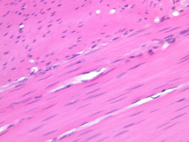
Skeletal muscle fibers differ in length, width, and the type of biochemical system for energy production. The two types of fibers also differ in contractile properties; one in fast twitch or white muscle or phasic (for sprinting) and the other is slow twitch or red muscle or tonic (for marathon running), which contains myoglobin. There is also an intermediate fiber type.
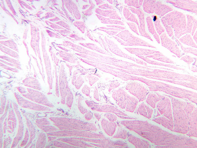
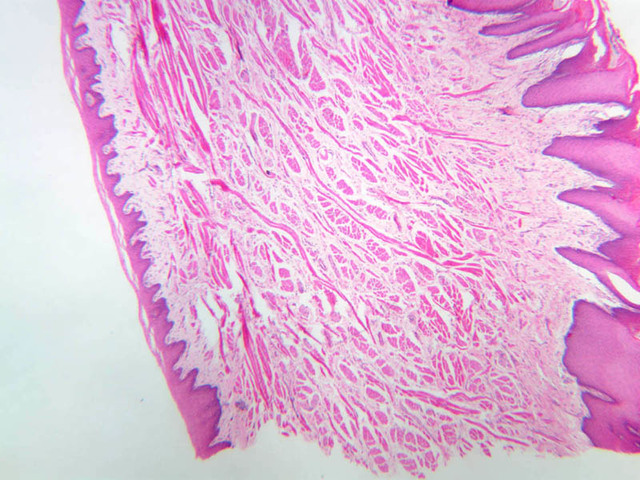
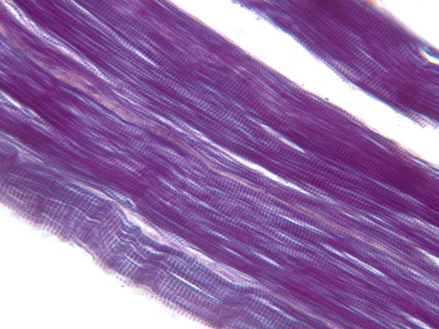
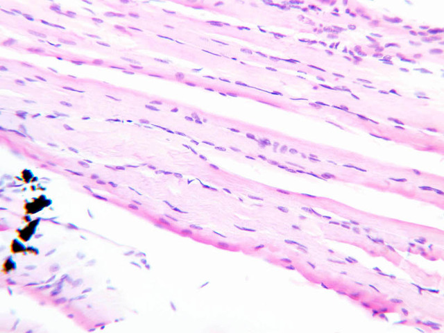
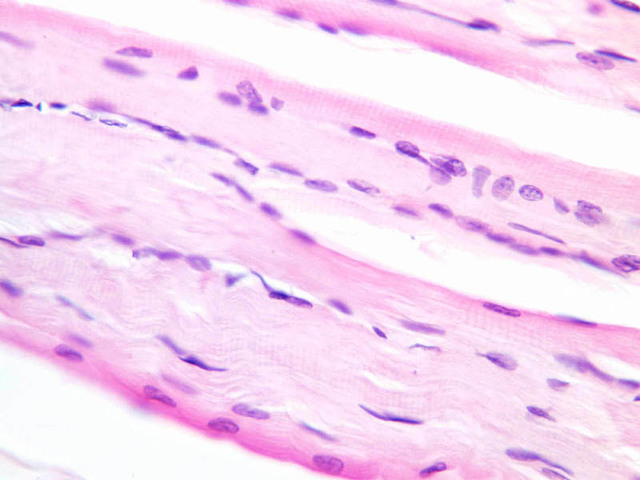
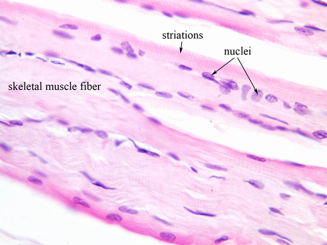
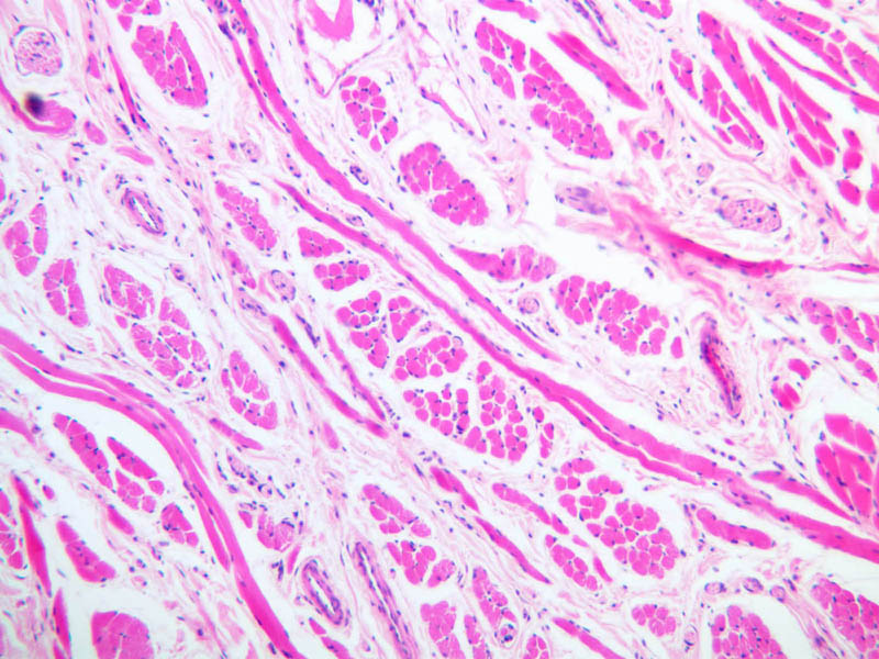
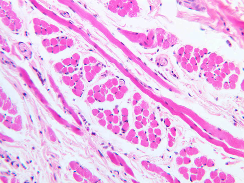
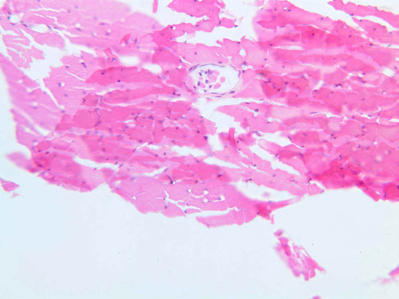
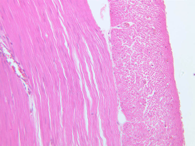
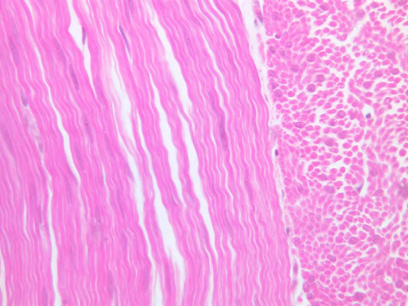
The striated skeletal muscle cell is an elongated, multinucleated cell, commonly called a muscle fiber. Most fibers are between 10 and 100μ wide and a few millimeters to a few centimeters long. Individual fibers of striated muscle (slide A-62 [
2.5x, 10x,
20x,
40x-labeled]; slide A-64 [
2.5x,
10x,
20x,
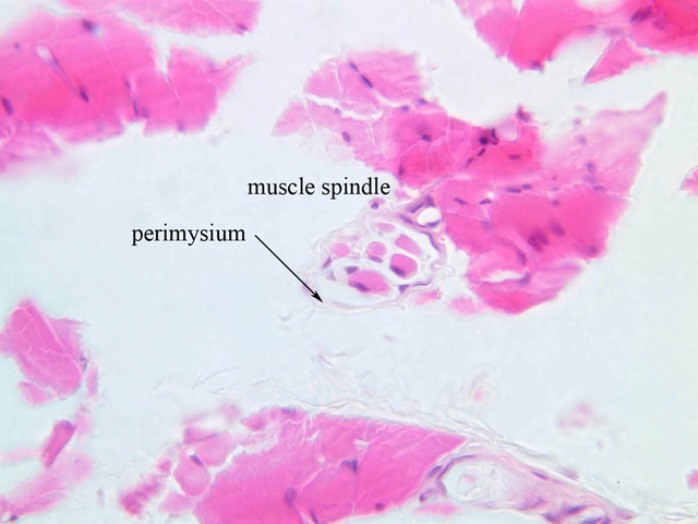
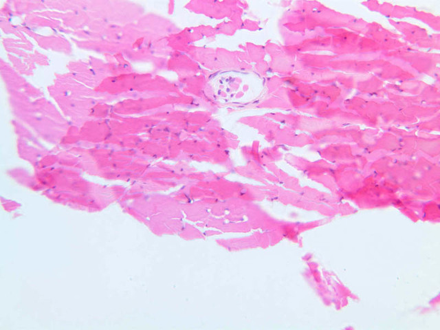
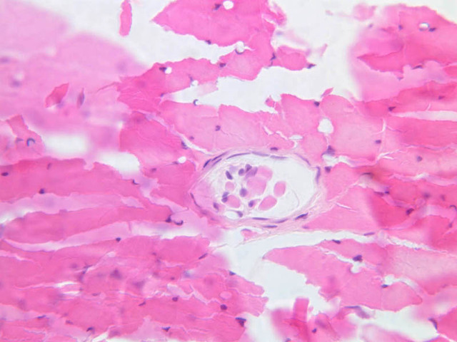
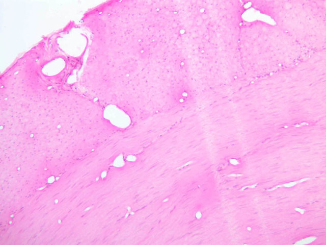
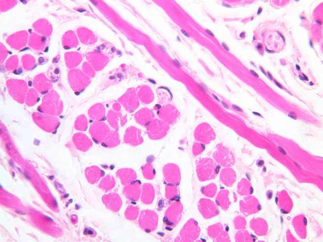
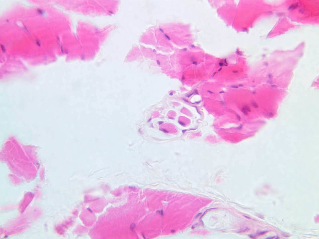
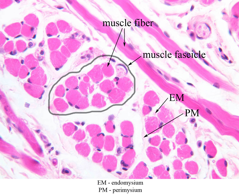
40x-labeled]) (seen in cross section) are sheathed by a delicate layer of connective tissue called endomysium. In the individual fibers, myofibrils, which appear in the section as dots, occur in irregular clumps separated by clear lines (of sarcoplasm). Nuclei are usually located around the periphery of skeletal fibers, but occasional examples of deeper-lying nuclei can be found. The density of nuclei in any one fiber is related to the size of the fiber. As in the nervous tissue, individual fibers are gathered into bundles, enclosed by connective tissue called perimysium in this case. If the whole muscle were visible on the slide, it would be seen to have an external investment of loose (fascial) connective tissue, which is called epimysium. Most large skeletal muscles are united to a tendon, which in turn is attached to a bone. Blood vessels penetrate the muscle through the various mysial layers of connective tissue and end in capillaries around individual fibers. Motor and sensory nerve fibers have a similar distribution. Sensory nerves are particularly associated with muscle spindles, which consist of several smaller-than-usual skeletal fibers gathered by perimysium into bundles. These structures, which serve as stretch receptor units, are present on slide A-91 (lumbricals [
10x,
20x,
40x-labeled] [
20x,
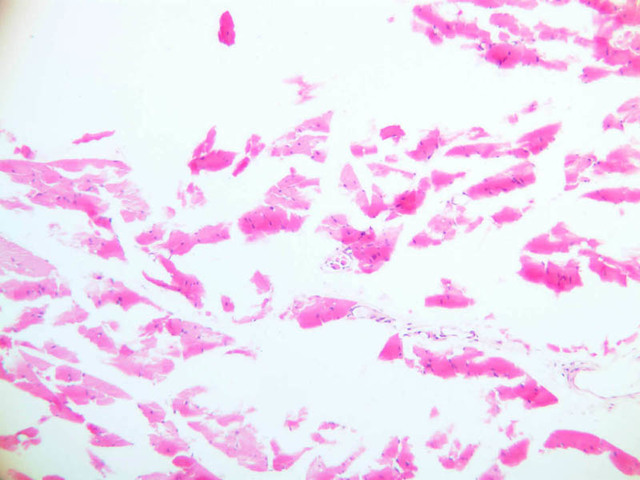
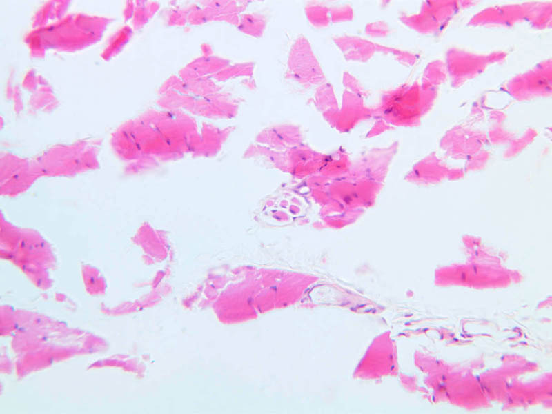
40x]). Locate an area of the section in which the striated muscle is cut longitudinally. Note the connective tissue endomysium and numerous small blood vessels between individual muscle fibers. Look for groupings of fibers that form fascicles, surrounded by coarse bands of perimysial connective tissue.
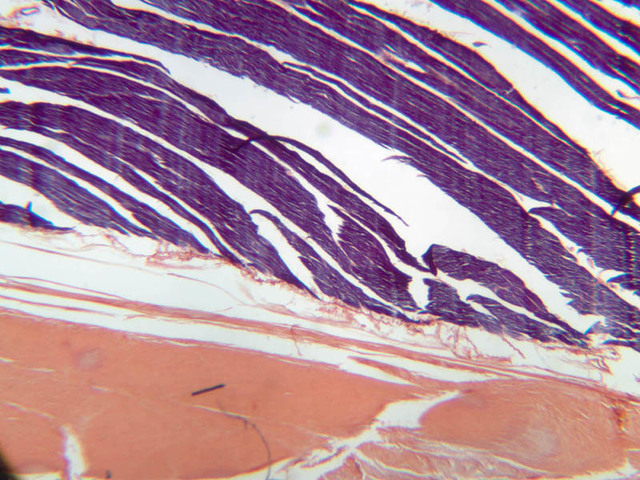
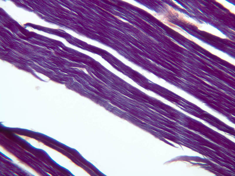
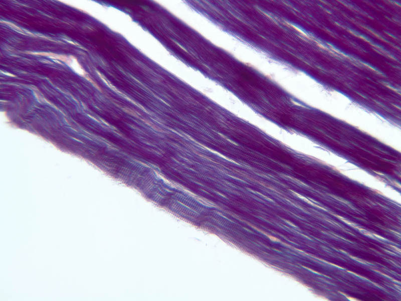
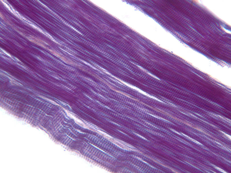
By light microscopy there are alternating bands of varying eosinophilia as in A-62 or A-64 and these can be differentially stained with dyes such as phosphotungstic acid hematoxylin or PTAH as in slide A-19 (PTAH [
2.5x,
10x,
20x,
40x]). Attempt to identify the boundary or cell membrane (in muscle called the sarcolemma) of the muscle fiber and note the peripheral location of the nuclei in each cell. Identify the alternating dark and less-dense bands extending from one to the other side of the fiber. The denser cross-bands are anisotropic in polarized light and from this characteristic are known as A bands. The less-dense cross bands are isotropic (i.e., do not rotate the plane of transmitted light) and are called I bands. The I band (composed of actin filaments) is bisected by the thin Z line (Ger., Zwischenband, bisecting line). That part of the myofibril that lies between two Z bands is called a sarcomere and is the contractile unit of the striated muscle cell.
The neuromuscular junction is best visualized at the EM level, but is also identifiable at the light microscopic level of resolution. Muscle cells have scattered surface invaginations referred to as primary synaptic clefts. It is in these clefts that the axon of the motor nerve terminates. Along the sides and base of the primary synaptic cleft are rather deep folds of the muscle cell sarcolemma. These folds are called secondary synaptic clefts or junctional folds. The contours of the membranes of the axon ending and muscle cell follow one another somewhat, but the nerve and muscle cell is always separated by a synaptic cleft containing the basement membrane surrounding the entire muscle. The muscle cell cytoplasm in the region of the neuromuscular junction has an abundance of mitochondria, rough endoplasmic reticulum and free ribosomes, perhaps for the synthesis of acetylcholine receptors in the membrane of the muscle cell. The axon innervating a neuromuscular junction lacks myelin. Instead, its associated Schwann cell forms a protective cap over the junction.
Skeletal Muscle Image Gallery
Skeletal Muscle Table of Identifications
| Row |
Structure |
Abbreviation |
Optimal Stain |
Representative Section |
Note |
| 1 |
Skeletal Muscle Fiber |
(none), arrows |
H&E |
 A62, Tongue, 40x; A62, Tongue, 40x;  A64, Tongue, 40x A64, Tongue, 40x |
|
| 2 |
Striations |
(arrow) |
H&E |
A62, Tongue, 40x |
|
| 3 |
Nuclei |
(arrows) |
H&E |
A62, Tongue, 40x |
|
| 4 |
Muscle Fascicle |
(outline) |
H&E |
A64, Tongue, 40x |
|
| 5 |
Endomysium |
EM |
H&E |
A64, Tongue, 40x |
|
| 6 |
Perimysium |
PM |
H&E |
A64, Tongue, 40x |
|
| 7 |
Muscle Spindle |
(none) |
H&E |
 A91, Lumbrical, 40x A91, Lumbrical, 40x |
|
Top of page
Cardiac Muscle
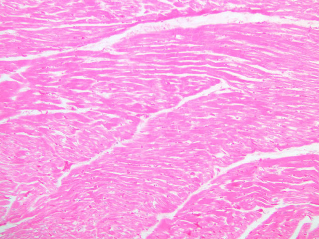
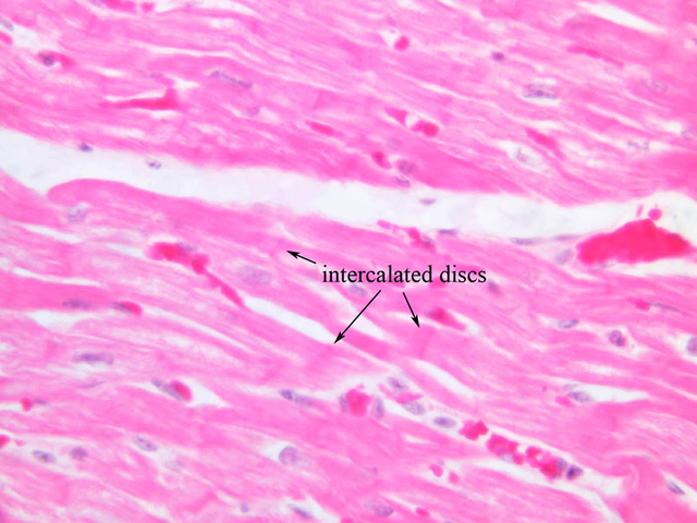
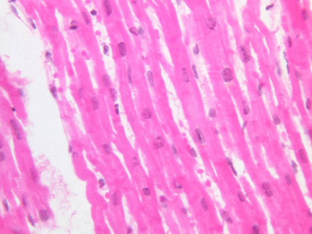
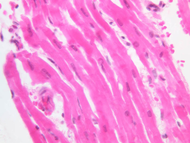
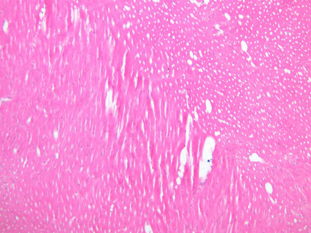
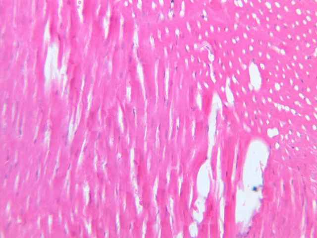
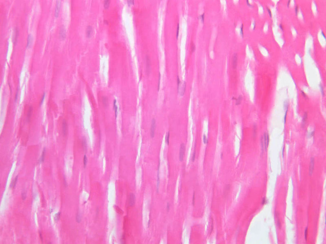
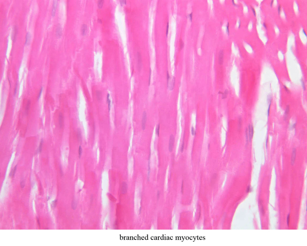
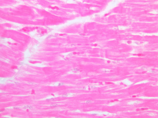
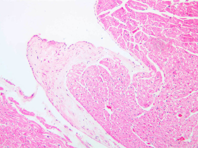
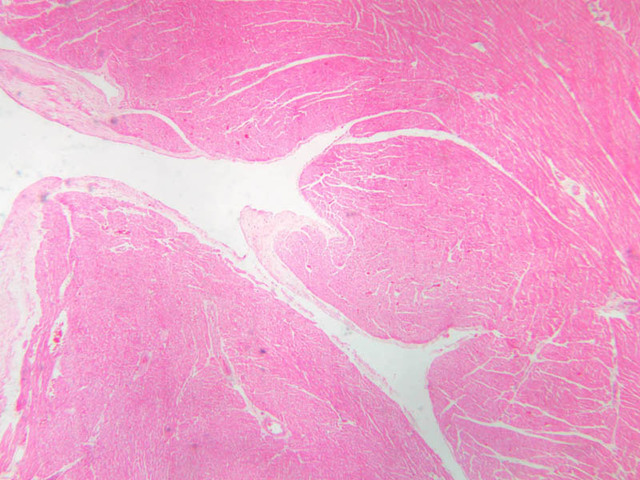
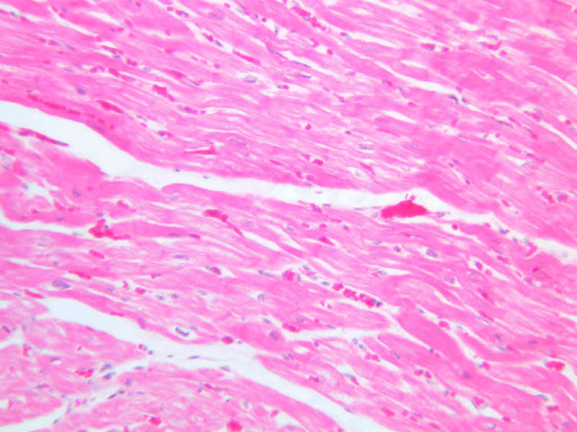
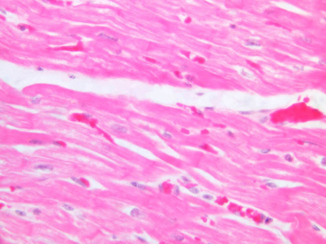
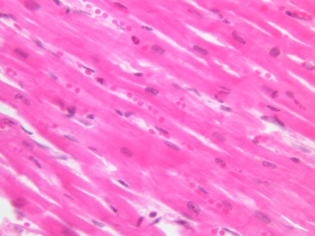
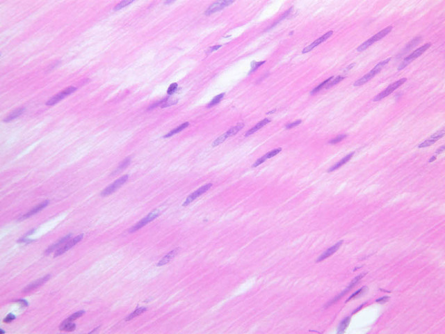
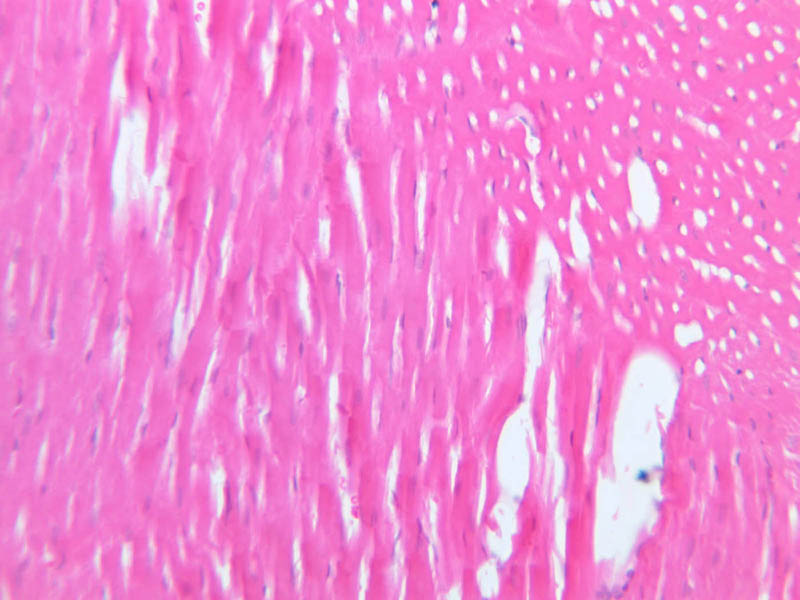
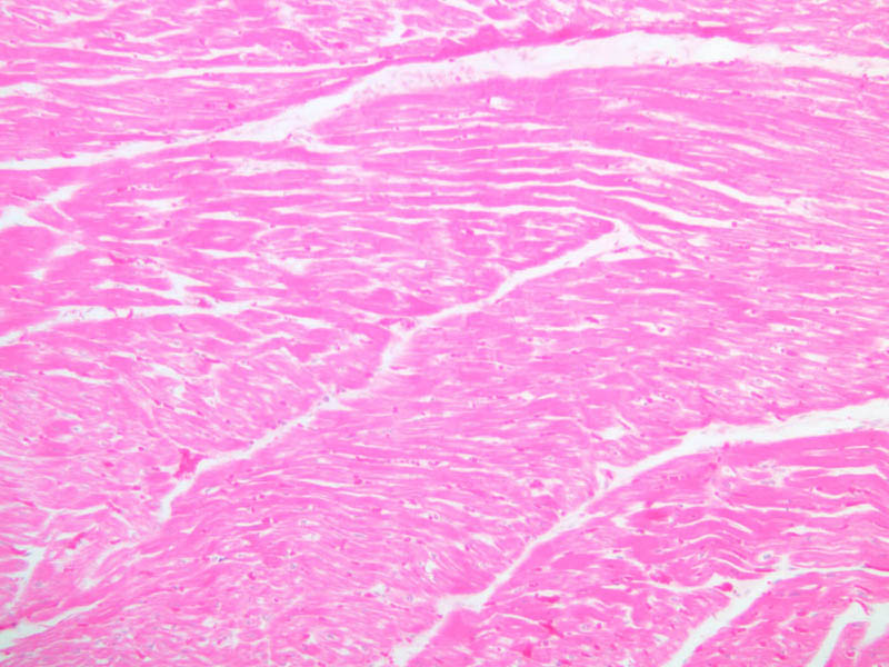
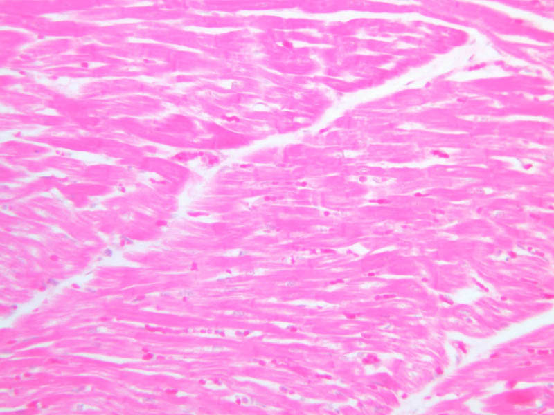
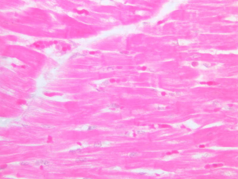
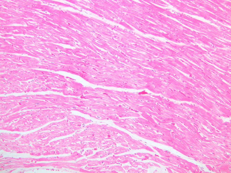
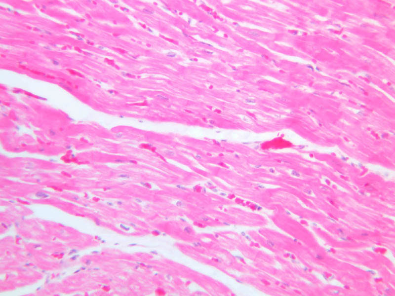
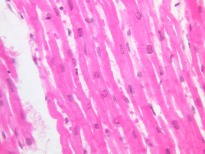
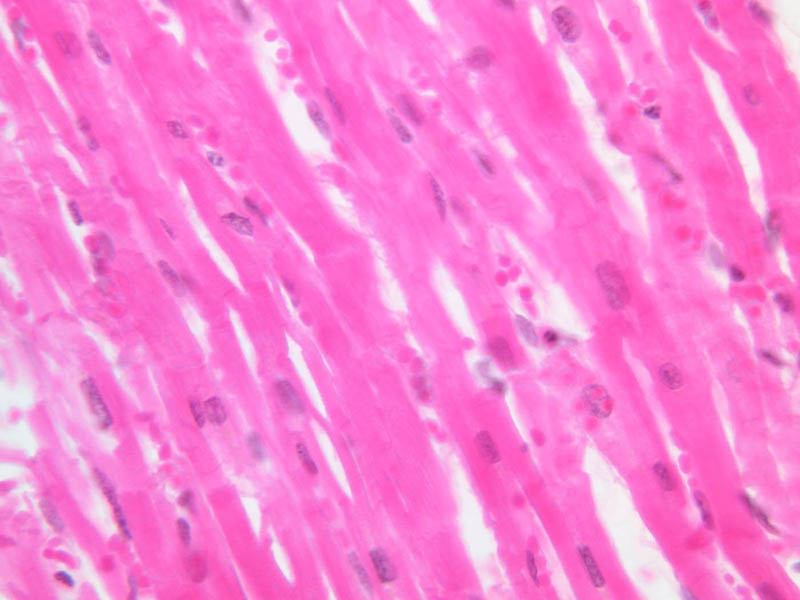
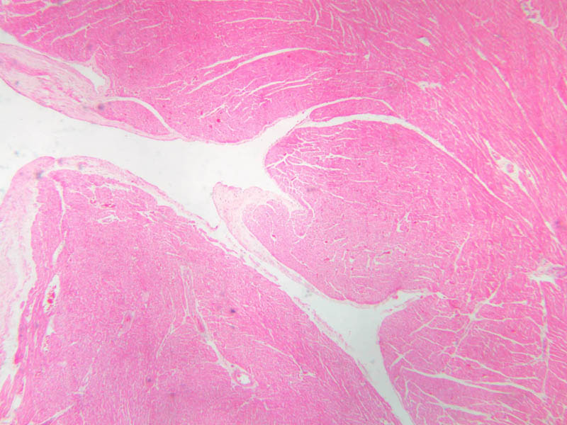
Cardiac muscle is also striated, but each muscle cell has a single nucleus and cells are attached one to the other by junctional complexes forming a structural as well as functional syncytium. The intercalated disc is the distinctive ultrastructural specialization in cardiac muscle often seen as a suggestion of a step-like density along the course of individual muscle cells. Intercalated discs are visible on slides A-20 ([
10x,
20x,
40x-labeled]) and A-25 ([
10x,
20x,
40x] [
10x,
20x,
40x-labeled] [
40x,
40x,
40x,
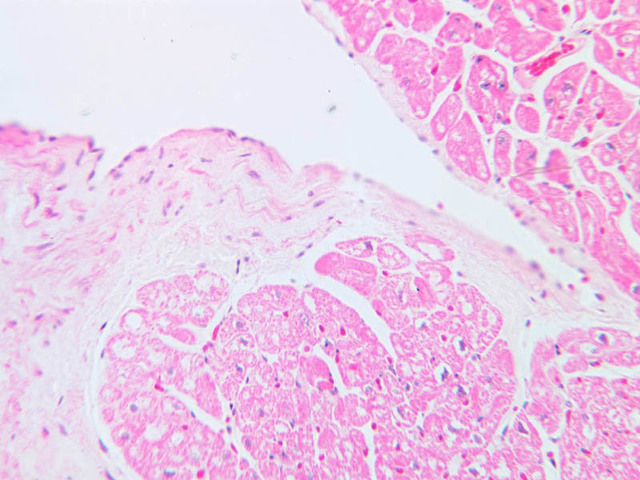
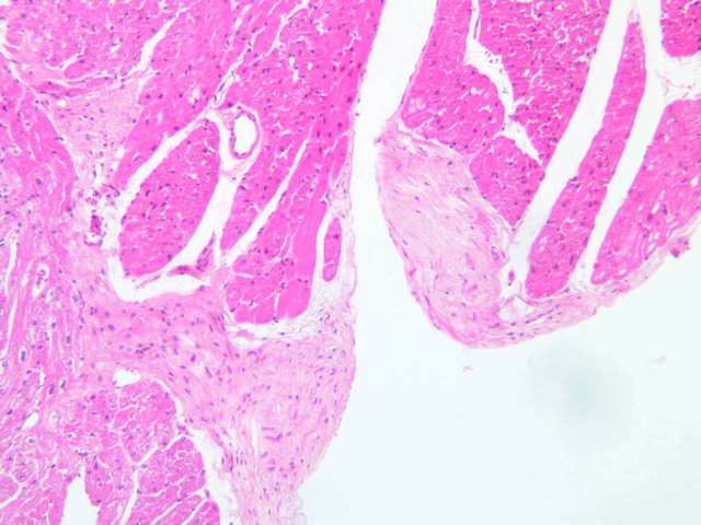
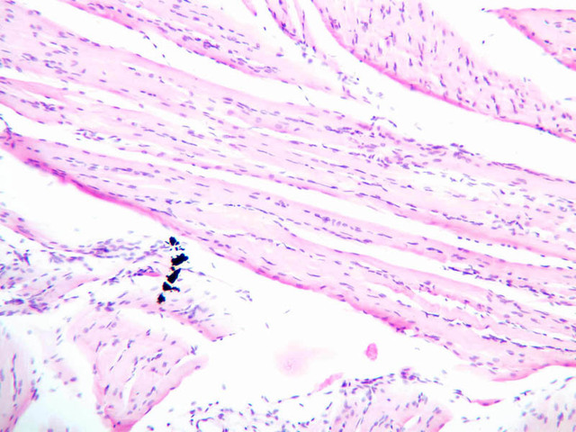
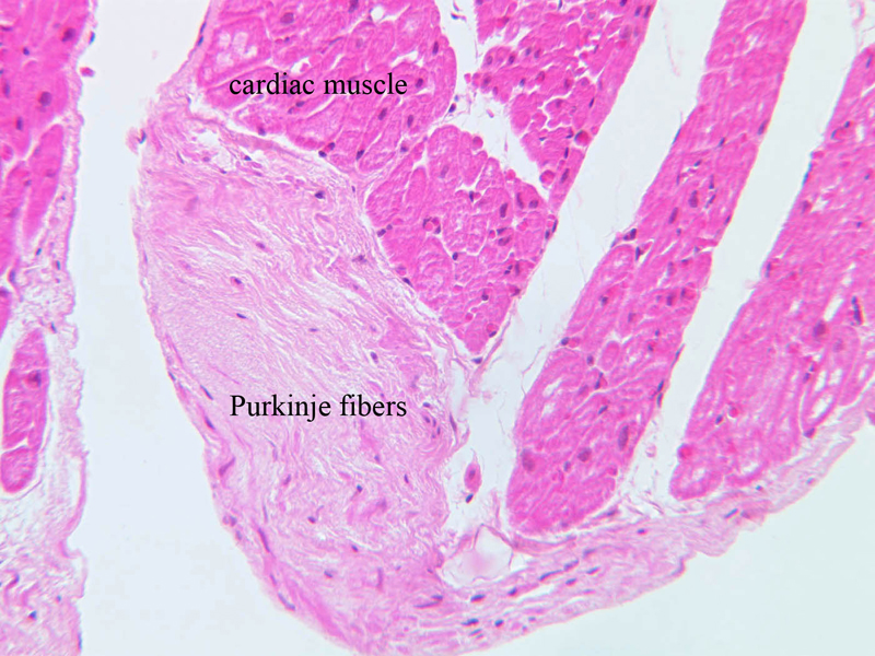
40x]). Cardiac muscle is distinctive by the branching pattern since each muscle cell has to attach to more than one other cardiac muscle cell giving rise to the appearance of a branched system. Cardiac fibers branch and interweave, and thus produce a squeezing action when the muscle contracts. Look for branched fibers. The central nuclei are also distinctive. The presence of gap junctions along the longitudinal components of the myofibers provide a functional coupling of adjacent cells, thus stimulating the cardiac tissue at any one point will result in a wave of excitation sweeping over the entire complex of myocardium. The transmission of the specialized conductive system in the heart from the atrium to the ventricles, i.e., the bundle of His and its associated Purkinje fibers, is important in terms of integrating the action of the two ventricles and pulling the apex of the heart toward the base. Examine slide A-25 for bundles Purkinje fibers ([
2.5x,
10x,
20x,
40x] [
10x,
20x-labeled,
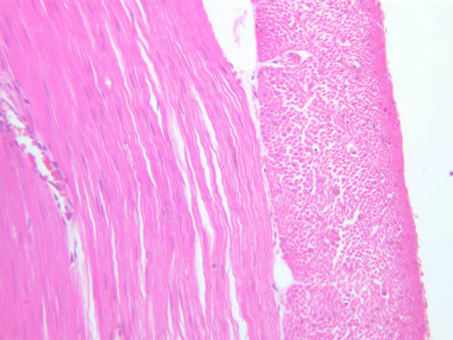
40x]). These specialized muscle cells will have a larger diameter than cardiac myocytes and they have lightly stained, eosinophilic cytoplasm. The central area of each cell appears especially light due the presence of glycogen.
Cardiac Muscle Image Gallery
Cardiac Muscle Table of Identifications
| Row |
Structure |
Abbreviation |
Optimal Stain |
Representative Section |
Note |
| 1 |
Branched Cardiac Myocytes |
(none) |
H&E |
 A20, Cardiac Muscle, 40x A20, Cardiac Muscle, 40x |
|
| 2 |
Intercalated Discs |
(arrows) |
H&E |
 A25, Left Ventricle, 40x A25, Left Ventricle, 40x |
|
| 3 |
Purkinje Fibers |
(none) |
H&E |
 A25, Purkinje Fibers (Cardiac Muscle), 20x A25, Purkinje Fibers (Cardiac Muscle), 20x |
|
| 4 |
Cardiac Muscle |
(none) |
H&E |
A25, Purkinje Fibers (Cardiac Muscle), 20x |
|
Top of page
Fine Structure of Skeletal and Cardiac Muscle Cells
(Future EM images), identify myofibrils, I, A, H, M, and Z bands as well as sarcoplasmic reticulum and triads. Visualize the appearance of a cross-section of the myofibril through the A band and through the I band. Which band changes in width when the muscle cell contracts? Study the three-dimensional arrangement of myofibrils, sarcoplasmic reticulum, and T-tubules.
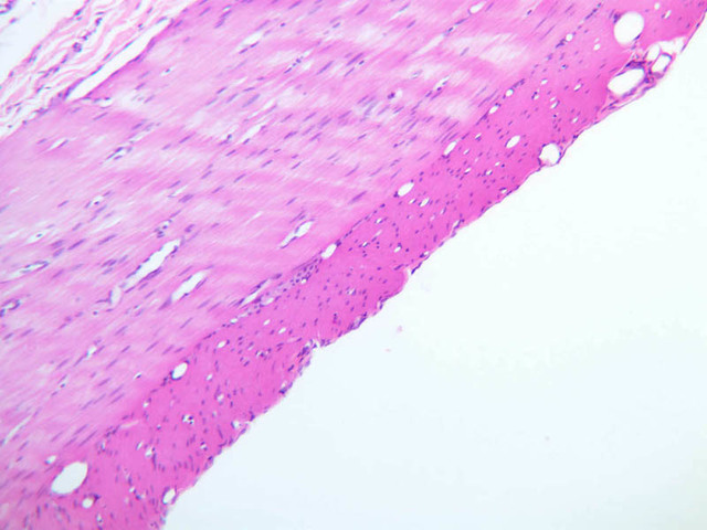
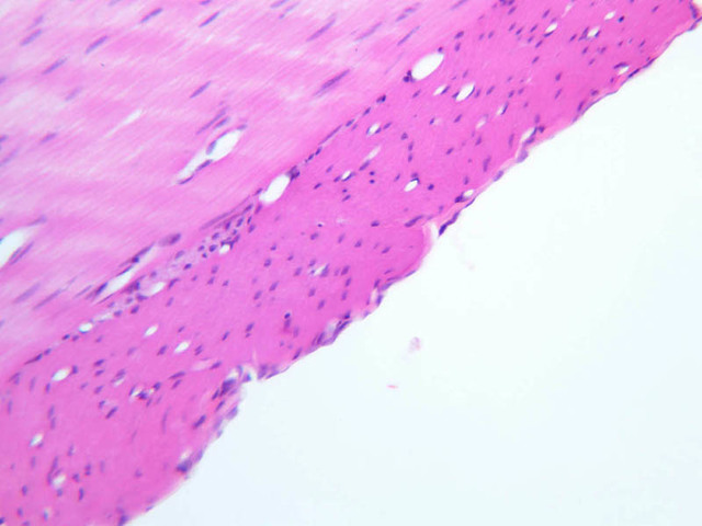
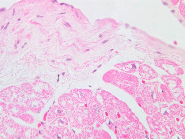
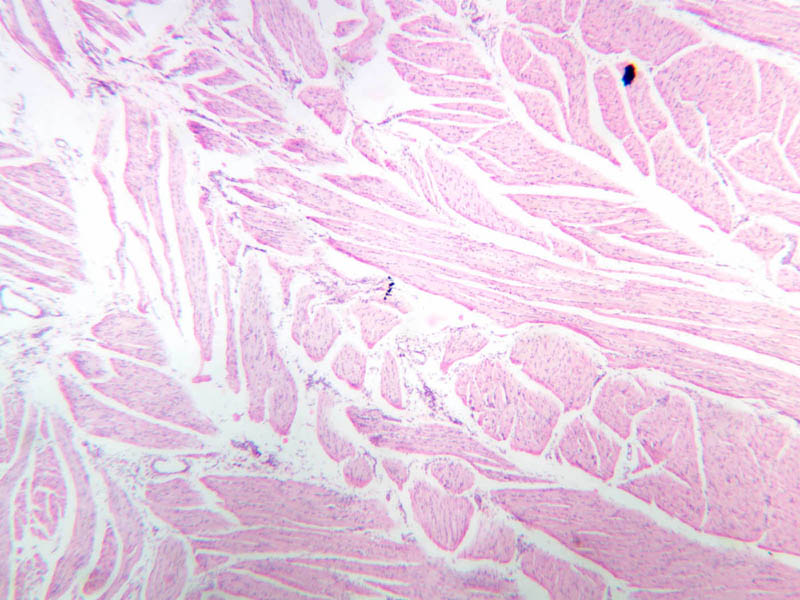
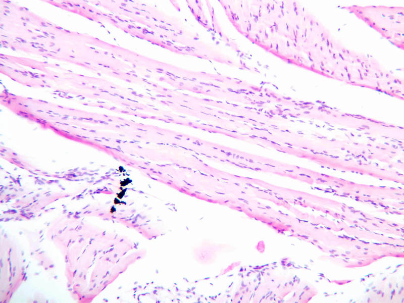
Smooth Muscle
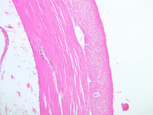
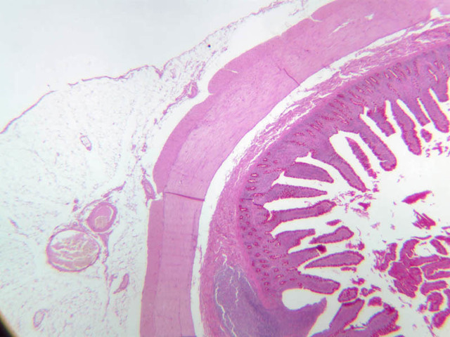
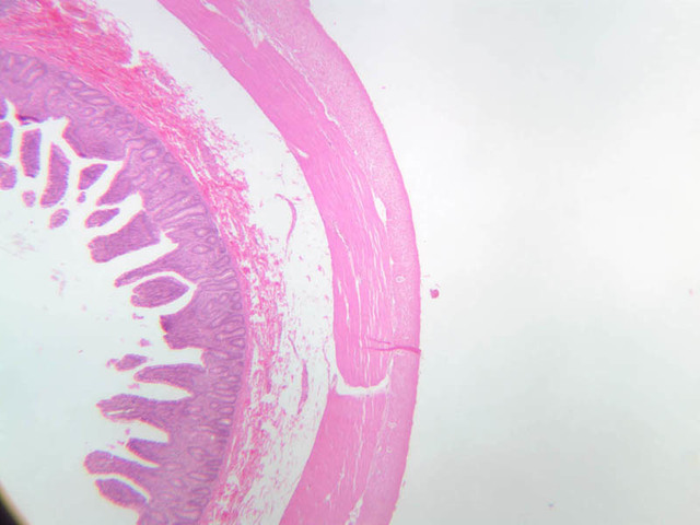
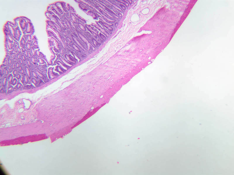
Smooth muscle is widely distributed throughout the body and it forms a component of vascular, renal, reproductive, and intestinal systems. Thus, any examples from your class sets that contain blood vessels or components of the G.I. or G.U. tract will contain smooth muscle.
Examine an H&E cross-section of intestine (slides B-12 [
2.5x,
10x,
20x,
40x] [
40x-labeled,
40x]; B-15 [
2.5x,
10x,
20x,
40x]; B-16 [
2.5x,
10x,
20x,
40x]; B-23 [
2.5x,
10x,
20x,
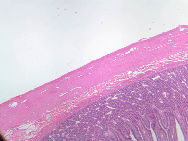
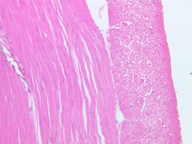
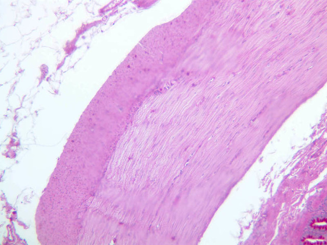
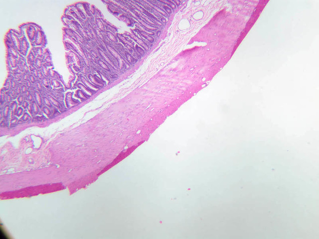
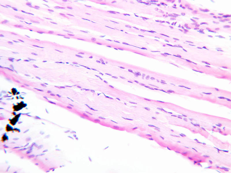
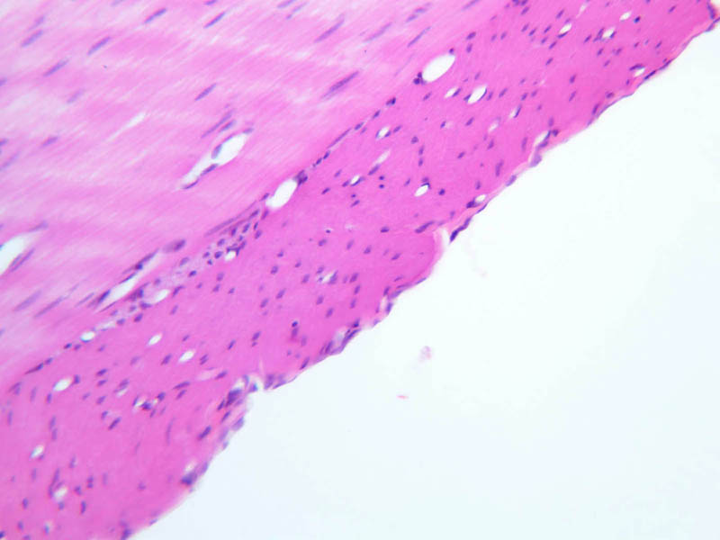
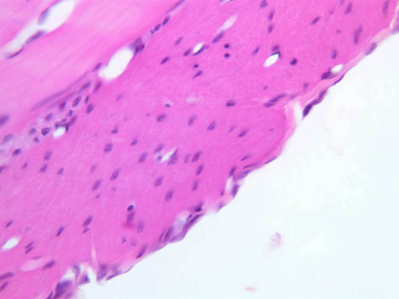
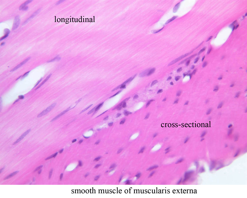
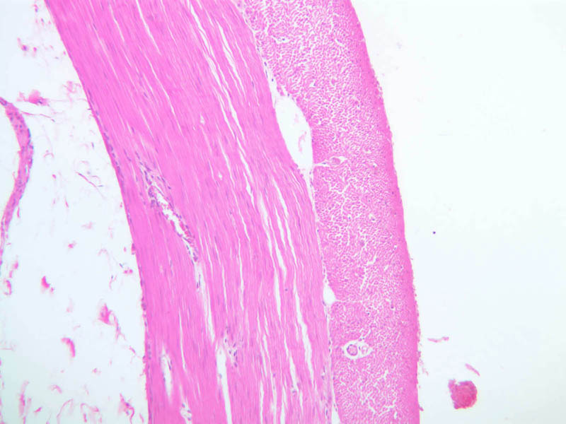
40x]). The smooth muscle occurs in a layer called the muscularis externa. The microscopic anatomy of the G.I. system will be dealt with later. Within the muscularis externa, there is an inner, circular layer and an outer, longitudinal layer.
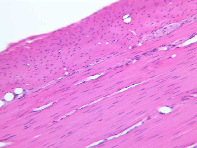
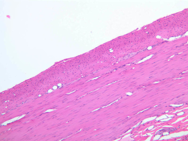
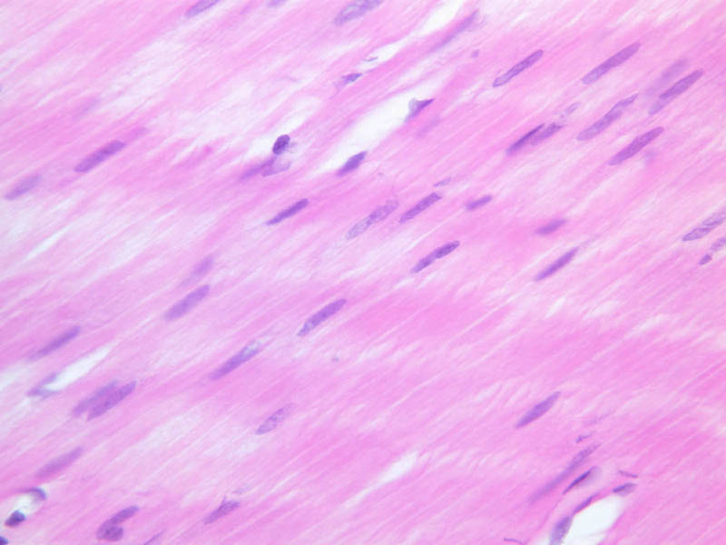
Study the muscle cells, which are sectioned longitudinally, in the inner layer. Attempt to follow the outlines of individual muscle fibers, and identify the nucleus in the center of the cell. Find a part of the section in which the cells of the outer longitudinal layer are cut most nearly in cross-section. Why does the nucleus not appear in all muscle cells seen in cross-section? The shape of the smooth muscle cell may be easier to see in sections of intestine stained by the PAS procedure (slides B-13 [
40x]; B-17 [
2.5x,
10x,
20x,
40x]; B-25 [
10x,
20x,
40x]) in which PAS-positive material outlines each muscle cell.
Study the fine structure of smooth muscle cells in (Future EM images). Note the distribution of the abundant filaments and the relative scarcity of other cytoplasmic structures such as endoplasmic reticulum. Identify the cytoplasmic densities that are thought to be homologous to the Z line in striated muscle cells and that apparently function to bind filaments together and to attach them to the plasma membrane.
Areas of direct connectivity of adjacent cells are referred to as gap junctions ("nexus"). Identify gap junctions in (Future EM images). These membrane specializations are points of low impedance and provide a direct electrical coupling between adjacent smooth muscle cells in order to provide synchronous activity of smooth muscle cells. Smooth muscle is innervated by nerves that are present at some distance from the muscle cell and the excitatory neurotransmitters will in turn diffuse through the connective tissue. Each smooth muscle cell is surrounded by basal lamina-like material as noted in the above electron micrographs.
Smooth Muscle Image Gallery
Smooth Muscle Table of Identifications
Top of page
Chapter Three Review
Review of Slides
Review of Identifications
| Row |
Structure |
Abbreviation |
Optimal Stain |
Representative Section |
Note |
| 1 |
Skeletal Muscle Fiber |
(none), arrows |
H&E |
A62, Tongue, 40x; A64, Tongue, 40x |
|
| 2 |
Striations |
(arrow) |
H&E |
A62, Tongue, 40x |
|
| 3 |
Nuclei |
(arrows) |
H&E |
A62, Tongue, 40x |
|
| 4 |
Muscle Fascicle |
(outline) |
H&E |
A64, Tongue, 40x |
|
| 5 |
Endomysium |
EM |
H&E |
A64, Tongue, 40x |
|
| 6 |
Perimysium |
PM |
H&E |
A64, Tongue, 40x |
|
| 7 |
Muscle Spindle |
(none) |
H&E |
A91, Lumbrical, 40x |
|
| 8 |
Branched Cardiac Myocytes |
(none) |
H&E |
A20, Cardiac Muscle, 40x |
|
| 9 |
Intercalated Discs |
(arrows) |
H&E |
A25, Left Ventricle, 40x |
|
| 10 |
Purkinje Fibers |
(none) |
H&E |
A25, Purkinje Fibers (Cardiac Muscle), 20x |
|
| 11 |
Cardiac Muscle |
(none) |
H&E |
A25, Purkinje Fibers (Cardiac Muscle), 20x |
|
| 12 |
Longitudinal Smooth Muscle of Muscularis Externa |
(none) |
H&E |
 B12, Jejunum, 40x B12, Jejunum, 40x |
|
| 13 |
Cross-sectional Smooth Muscle of Muscularis Externa |
(none) |
H&E |
B12, Jejunum, 40x |
|
Features for Histological Differentiation of Muscle Tissue
From, Sobotta/Hammersen Histology, Frithjof Hammerson, ed. Third ed., Lea & Febiger, Philadelphia, 1985.
Top of page
Top of page
--
AshleyLPistorio - 27 May 2007


