 |
|
You are here: Medical Histology>Main Web>AtlasContents>OralCavityAndSalivaryGlandsAtlas11 (20 Jun 2015, LorenEvey)Edit Attach
Chapter Eleven: Oral Cavity and Salivary Glands


Introduction
Prior to learning the microarchitecture of the Oral Cavity and Salivary Glands, use the table below to review some of the gross anatomy of these tissues:| Structure | Image |
|---|---|
| The Mouth | |
| The Oral Cavity and Pharynx | |
| Salivary Glands | |
| The Epiglottis | |
Oral Cavity
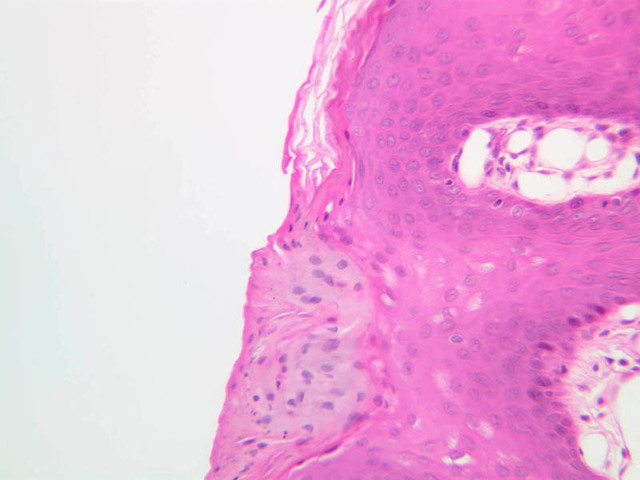
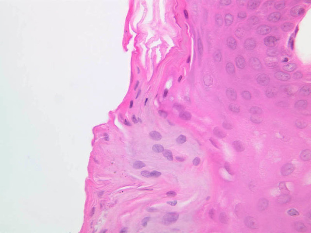
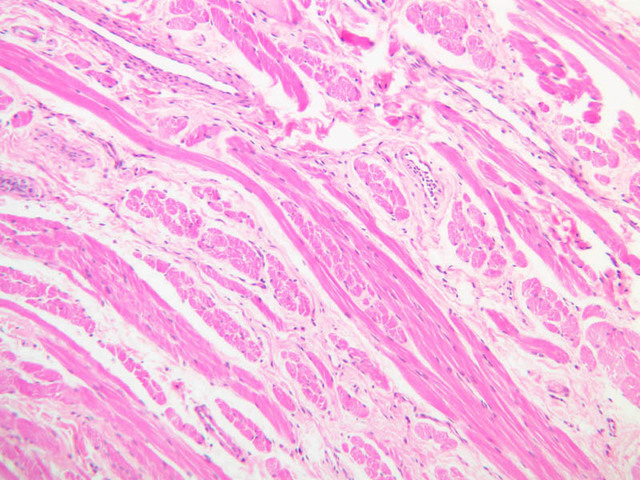
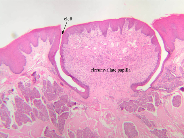
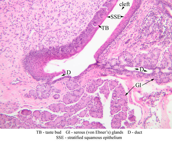
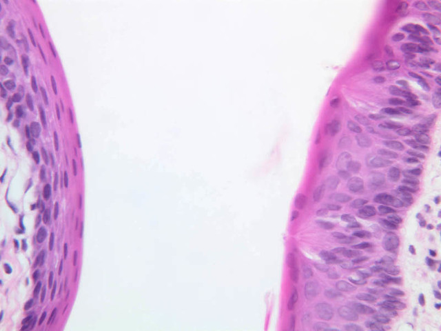
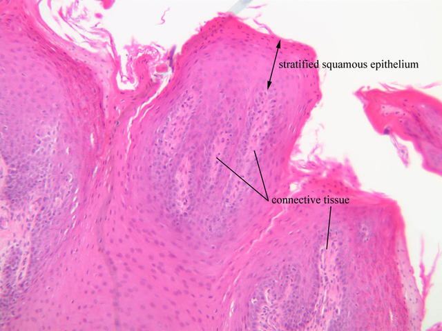
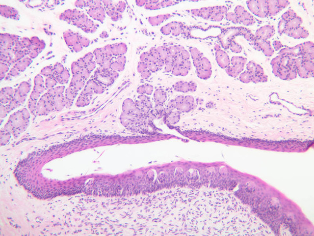
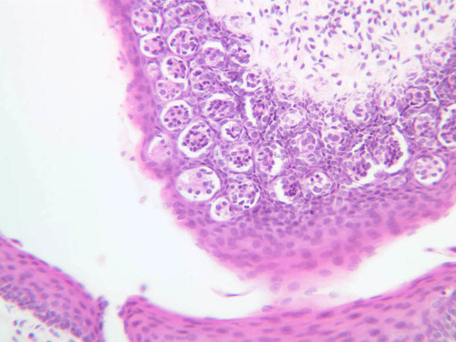
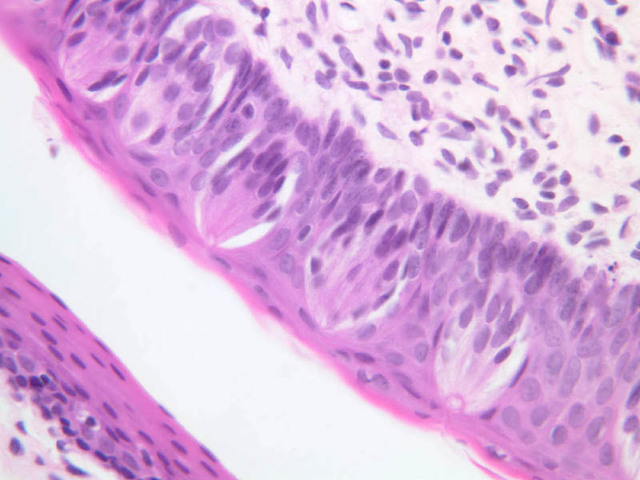
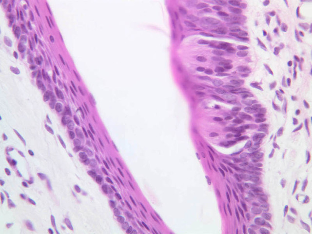
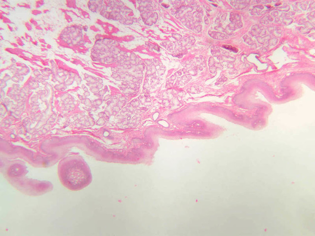
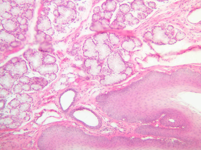
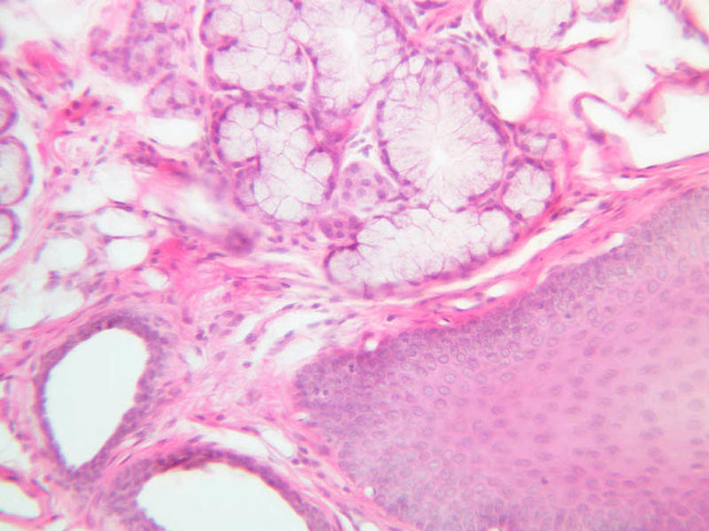
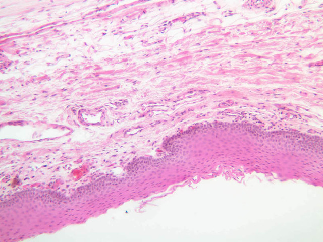
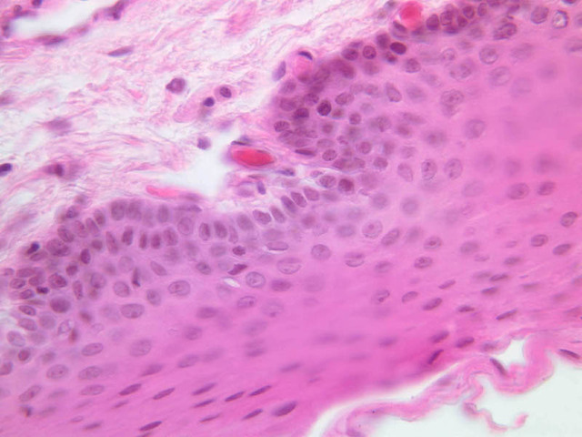
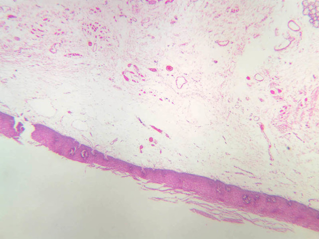
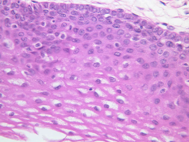
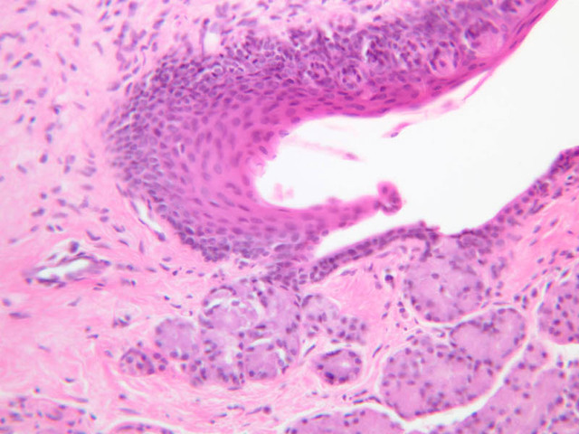
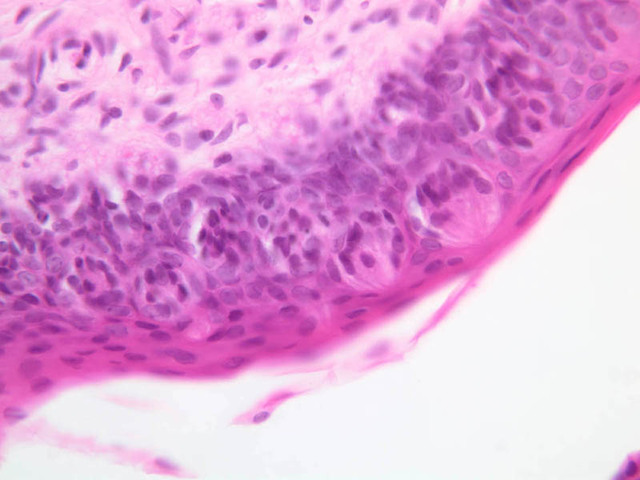
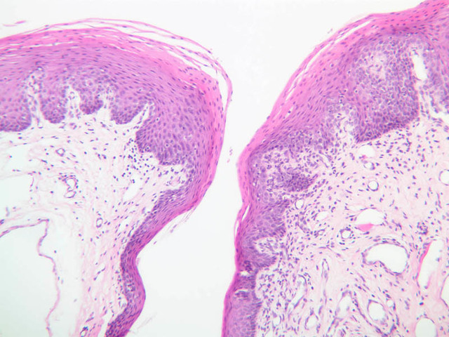
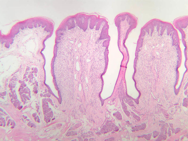
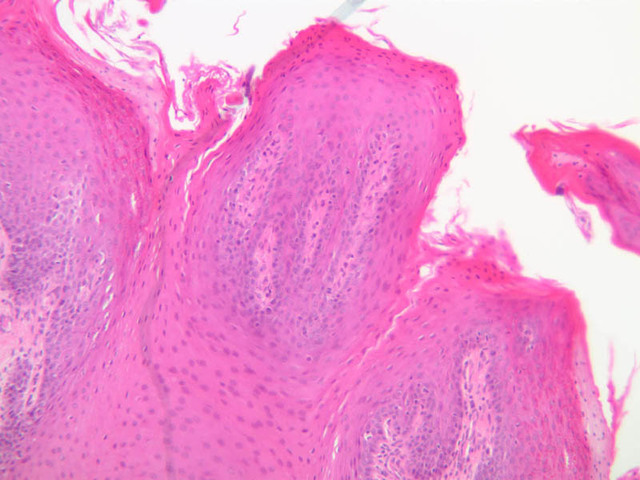
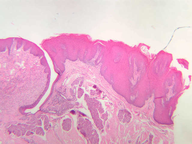
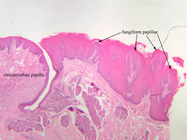
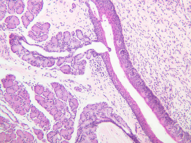
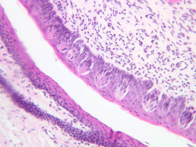
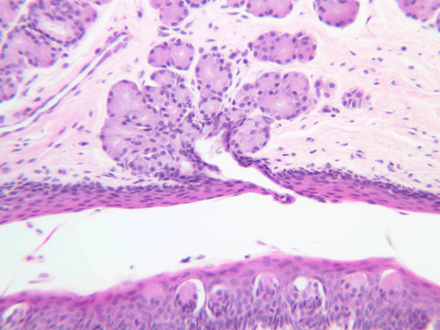
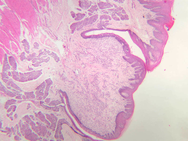
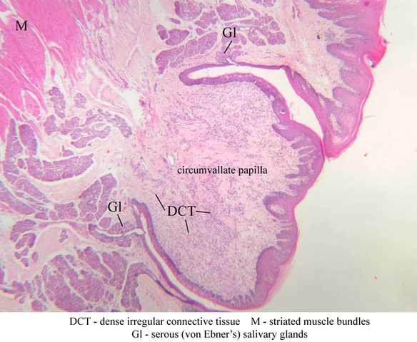
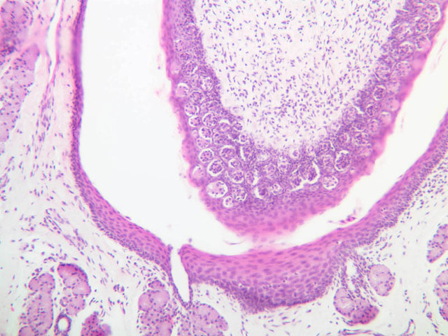
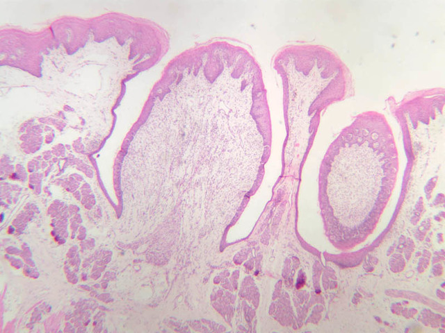
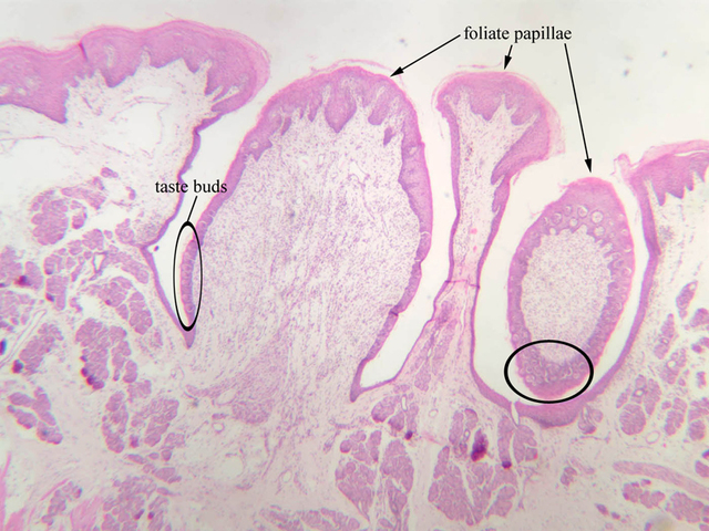
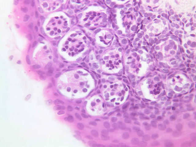
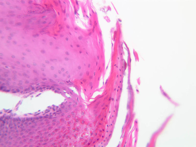
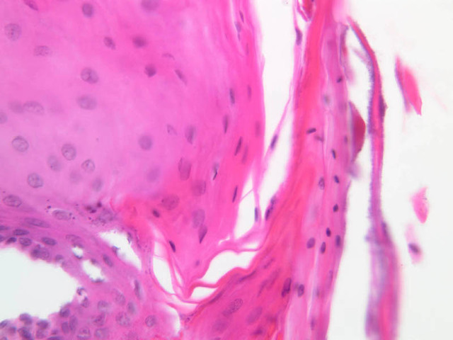
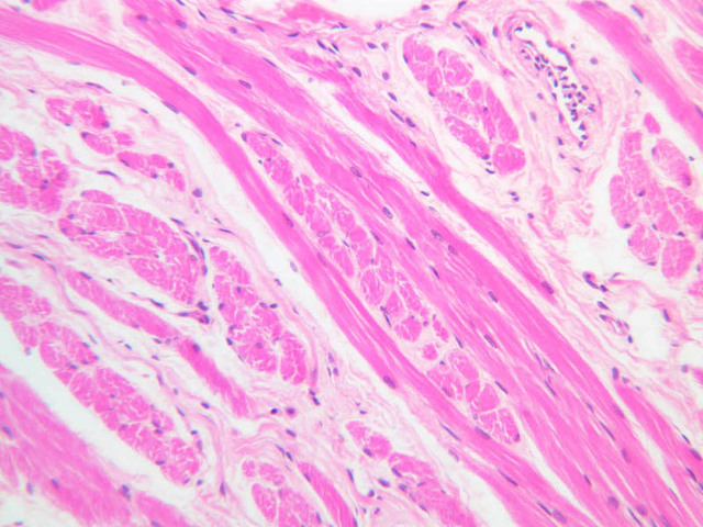
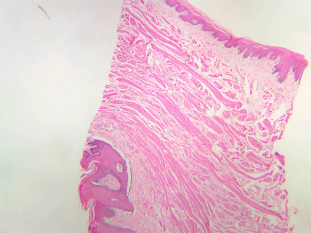
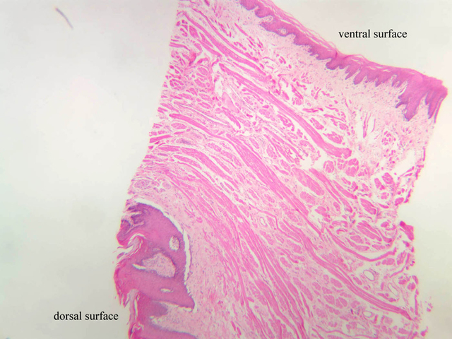
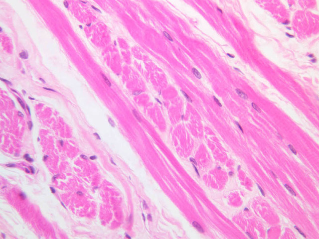
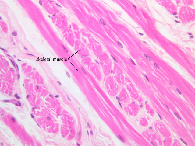
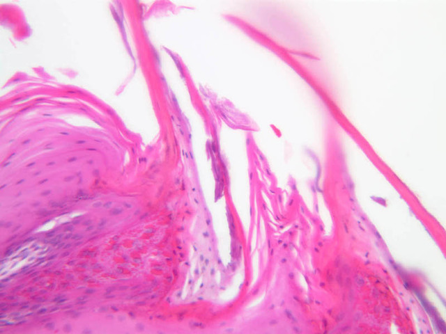
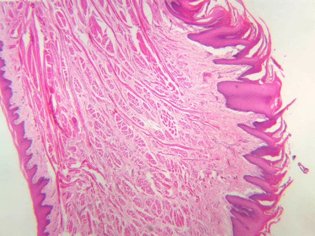
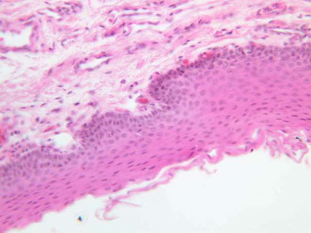
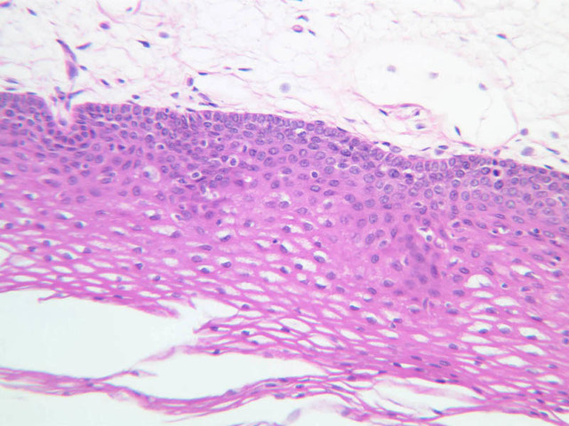
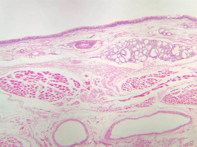
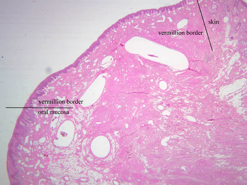
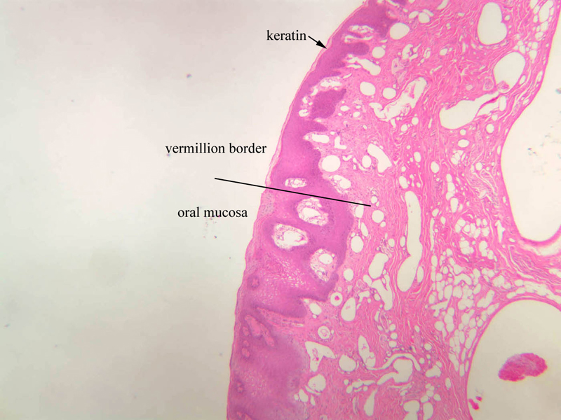
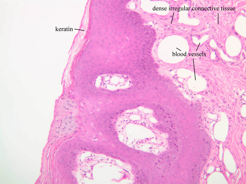
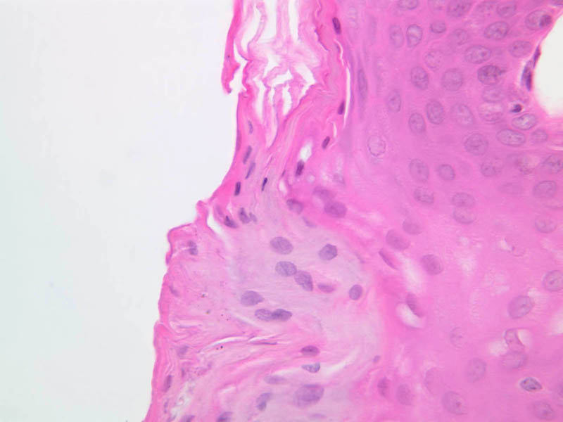
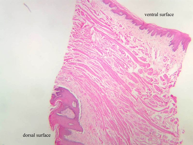
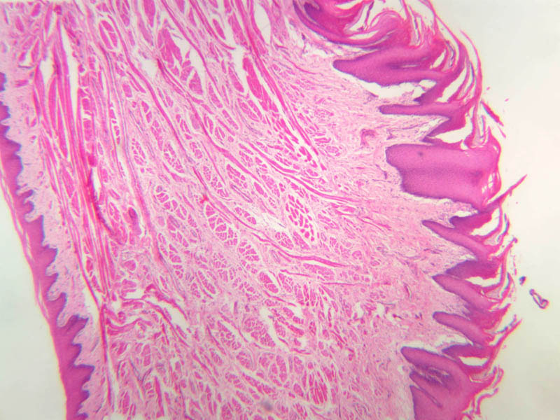
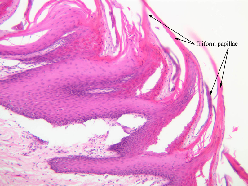
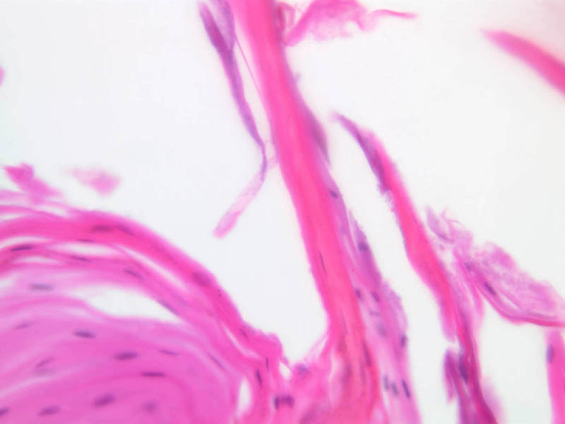
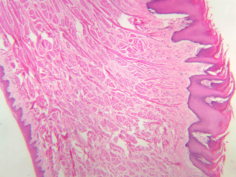
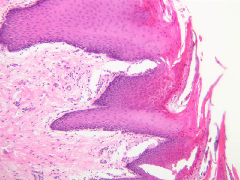
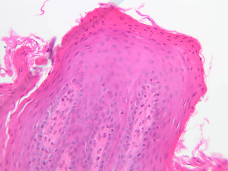
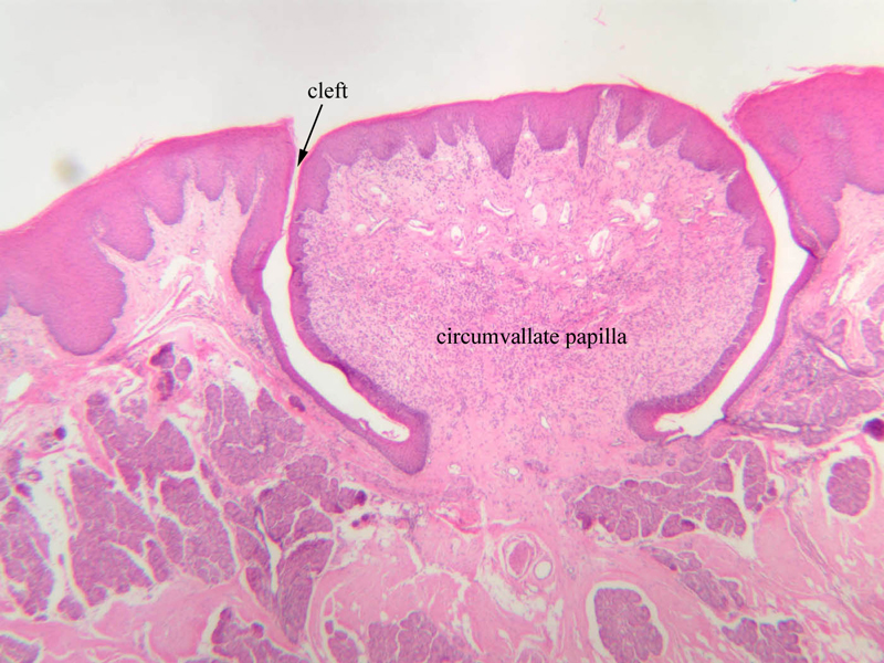
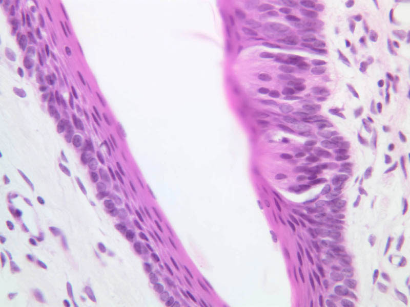
Using the lowest power objective, scan the section of human lip on slide A-55 [1x-labeled, 2x-labeled, 10x-labeled, 20x, 40x]. Note that the oral surface of the lip is covered by a very thick stratified squamous epithelium. Over the vermilion border (in life, the reddish portion) of the lip the stratified squamous epithelium is thinner and its outer layers are keratinized. This stratified squamous keratinized epithelium continues onto the outer surface of the lip, where sweat glands and hair follicles with their associated sebaceous glands appear in the underlying connective tissue bed. Note the abrupt change in epithelial thickness at the junction between the vermillion border and the oral mucosa; note also that this change in epithelial thickness pretty much coincides with a transition from keratinized to parakeratinized to nonkeratinized superficial layers. Look at your own lip and try to correlate what you see in the slide with what you see in the mirror. Underlying the epithelium is irregular CT. There is little distinction between the lamina propria and the submucosa. Blood vessels that lie just under the epithelium give lips their red color. Mucous glands (described below) can be found in the lamina propria on the mucosal side of the lip. Large masses of skeletal muscle are found in the lip. These represent slips of the orbicularis oris muscle. What nerve innervates this and the other muscles of facial expression? The tongue is covered with stratified squamous epithelium that, for the most part, is nonkeratinized. Whereas the ventral surface of the tongue is smooth, the dorsal and parts of the lateral surfaces are marked by alternating ridges and grooves that define the lingual papillae. Each papilla has a core of well-vascularized connective tissue that is continuous with the connective tissue that underlies all parts of the epithelium. The main mass of the tongue lies deep to this layer of connective tissue and consists of bundles of skeletal muscle that are oriented transversely, longitudinally and vertically. Using the section on slide A-64 [2.5x-labeled, 10x, 20x, 40x-labeled], examine the muscular core of the tongue and compare the mucosae of the dorsal and ventral surfaces. You should be able to find good examples of filiform papillae, which, as a rule, are the only parts of the tongue's surface that bear keratinized epithelium (A-63 [2.5x, 10x-labeled, 20x, 40x], [2.5x, 10x, 20x, 40x]. How does the epithelium of the inferior surface of the tongue differ from that of the superior surface? Your slide collection also has examples of fungiform (A-63 [2.5x-labeled, 10x-labeled, 20x, 40x] and foliate (A-63 [2.5x, 10x, 20x, 40x] papillae. Slide A-63 [2.5x-labeled, 10x-labeled, 20x, 40x] is a section of tongue through a circumvallate (vallate) papilla. Each circumvallate papilla is surrounded by a cleft or trench. Near the base of the papilla you should be able to find examples of von Ebner's (serous) salivary glands; you may also find profiles of the ducts of von Ebner's glands, which empty into the basal portion of the cleft (A-63 [2.5x-labeled, 10x, 20x, 40x]. Mucous glands, which stain less intensely than serous glands, are also present in the tongue. Especially over the lower two-thirds of the papilla, and spanning the full thickness of its epithelium, you should find a number of lightly stained, oval structures. These structures, which themselves have an epithelioid appearance, are taste buds (A-63 [2.5x-labeled, 10x, 20x, 40x] [2.5x, 10x, 20x, 40x] [40x]. In suitably stained material, taste buds can be shown to consist of three different cell types. The section in your collection does not lend itself to the identification of these cell types; however, it is suitable for the documentation of the rapid turnover of taste bud cells, as evidenced by the ease with which mitotic figures can be found.Oral Cavity Image Gallery


Oral Cavity Table of Identifications
| Row | Structure | Abbreviation | Optimal Stain | Representative Section | Note |
|---|---|---|---|---|---|
| 1 | Vermillion Border | (none) | H&E | |
|
| 2 | Skin (of Lip) | (none) | H&E | |
|
| 3 | Oral Mucosa | (none) | H&E | |
|
| 4 | Keratin | (none) | H&E | |
|
| 5 | Dense Irregular Connective Tissue | (none) | H&E | |
|
| 6 | Blood Vessels | (none) | H&E | |
|
| 7 | Dorsal Surface (of Tongue) | (none) | H&E | |
|
| 8 | Ventral Surface (of Tongue) | (none) | H&E | |
|
| 9 | Skeletal (Striated) Muscle | (none), M | H&E | |
|
| 10 | Filiform Papillae | (none) | H&E | |
|
| 11 | Circumvallate Papilla | (none) | H&E | |
|
| 12 | Cleft | (none) | H&E | |
|
| 13 | Taste Bud | TB, (circled areas) | H&E | |
|
| 12 | Serous (von Ebner's) Glands | Gl | H&E | |
|
| 13 | Duct | D | H&E | |
|
| 14 | Stratified Squamous Epithelium | SSE | H&E | |
|
| 15 | Fungiform Papillae | (none) | H&E | |
|
| 16 | Serous (von Ebner's) Glands | Gl | H&E | |
|
| 17 | Foliate Papillae | (none) | H&E | |
Back to Top of Page
Salivary Glands
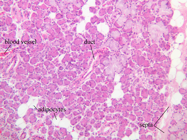
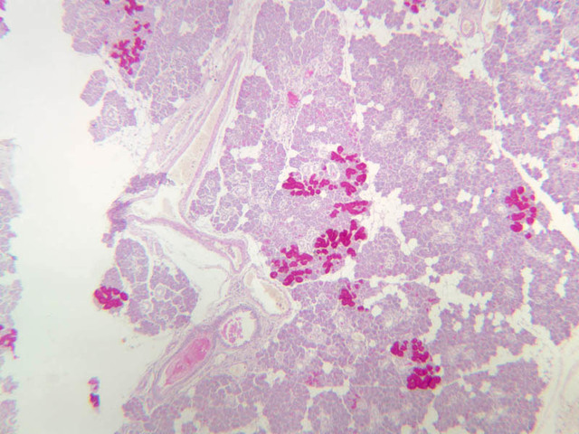
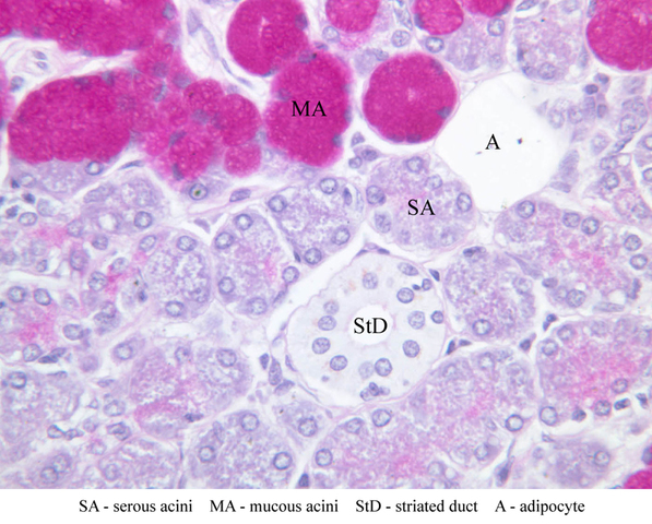
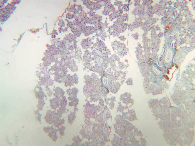
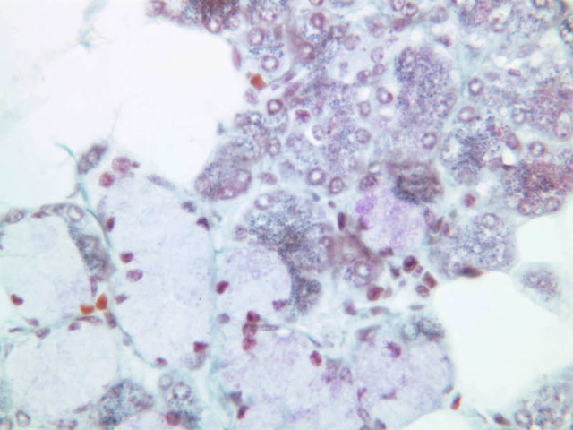
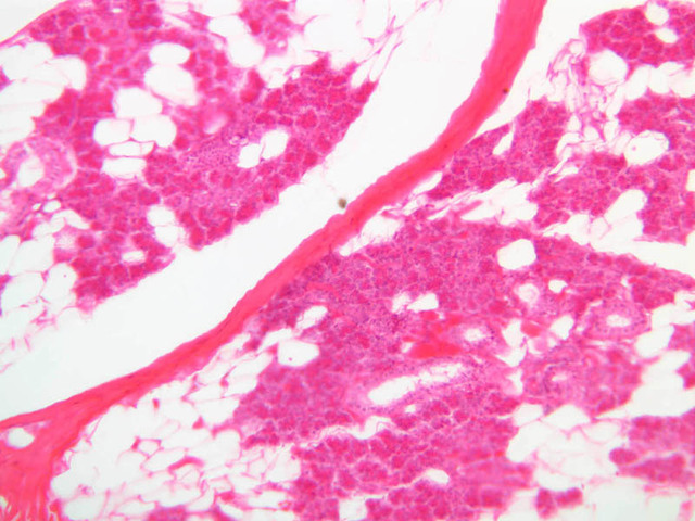
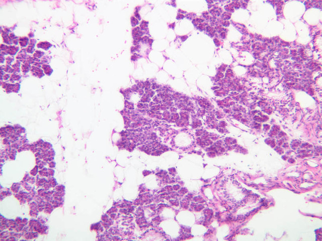
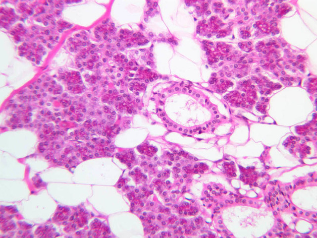
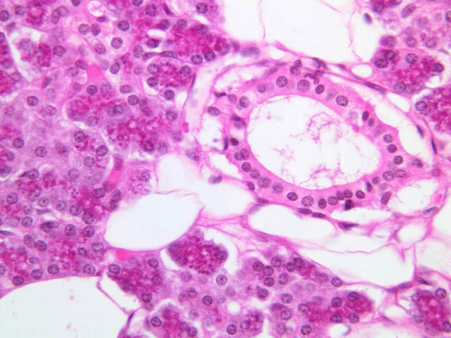
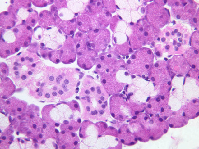
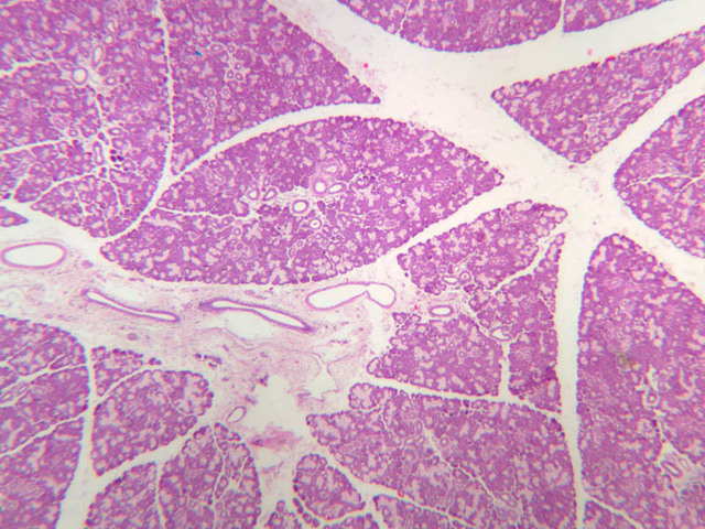
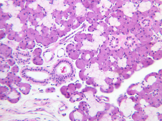
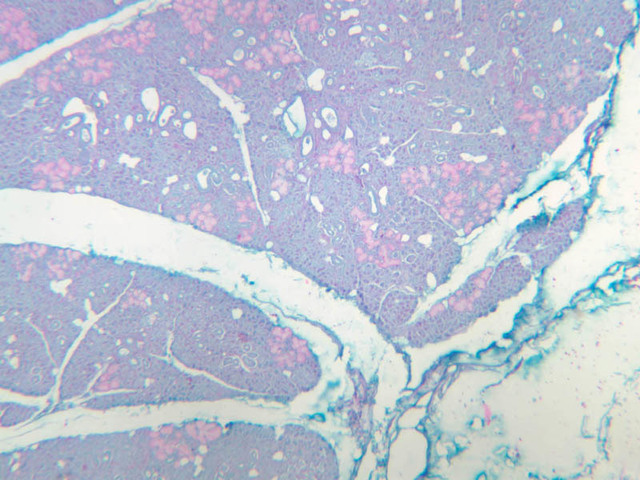
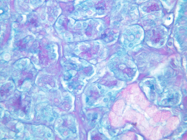
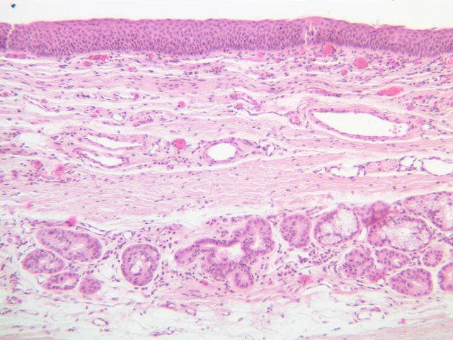
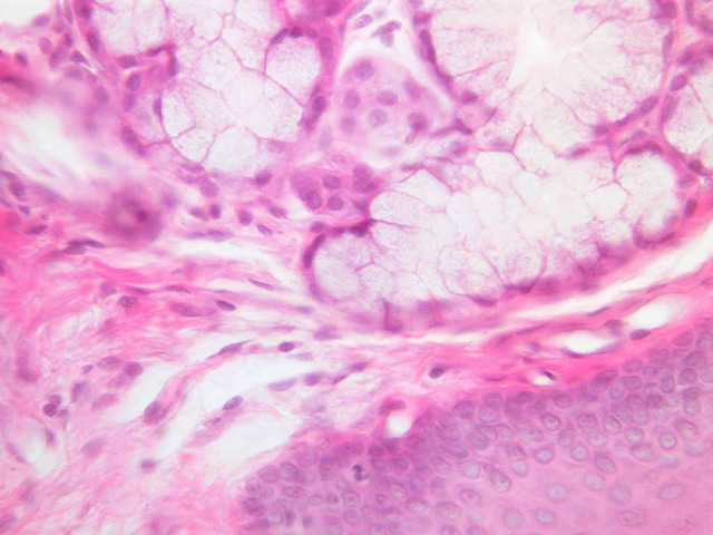
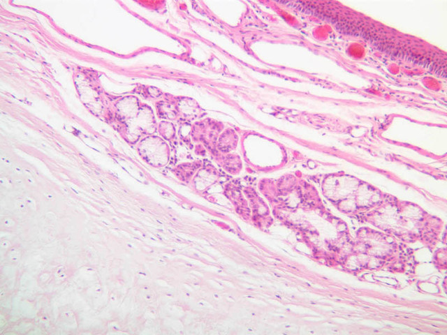
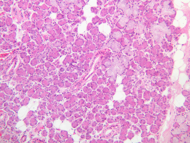
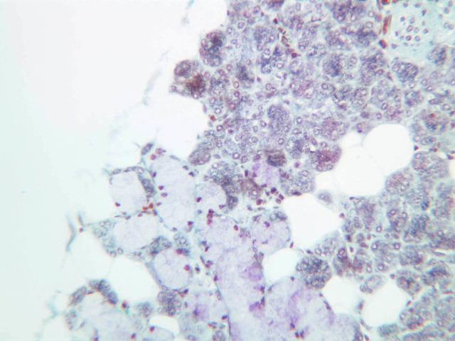
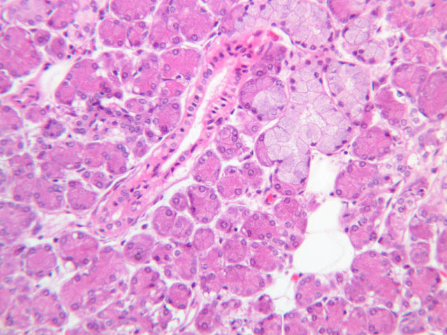
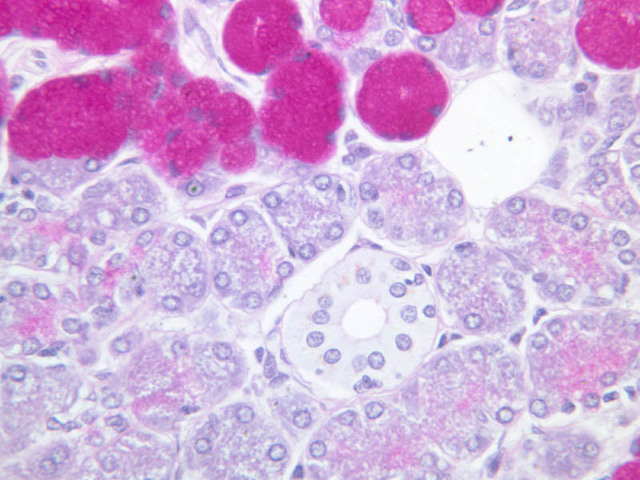
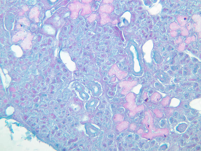
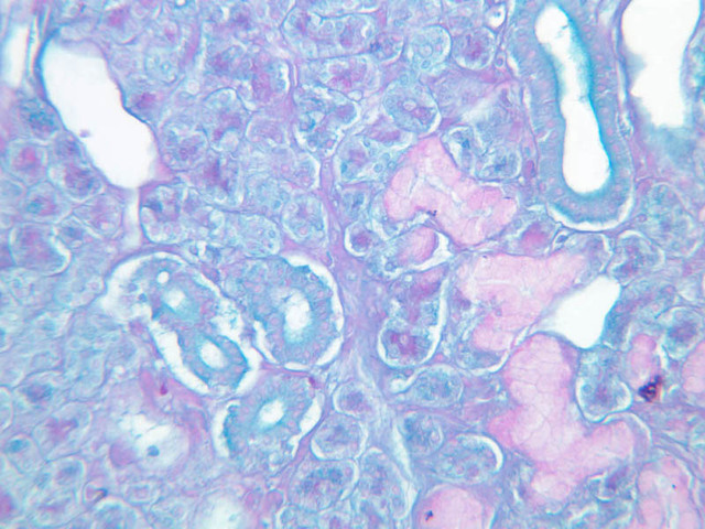
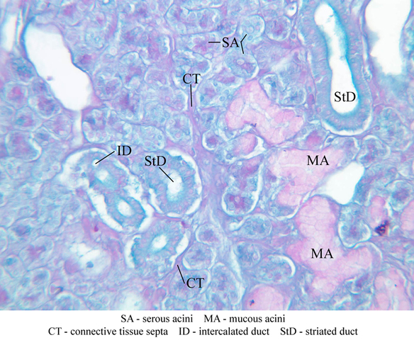
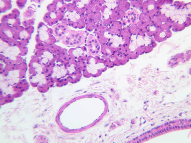
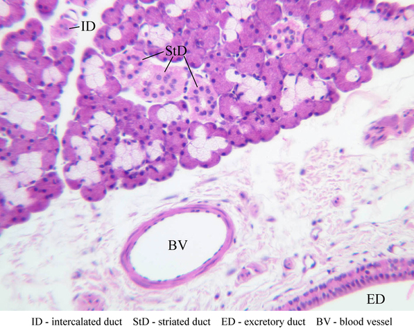
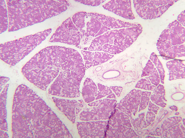
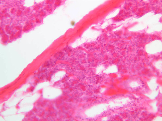
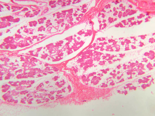
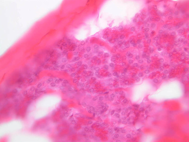
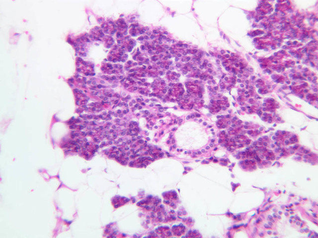
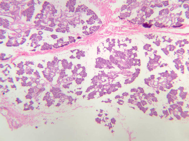
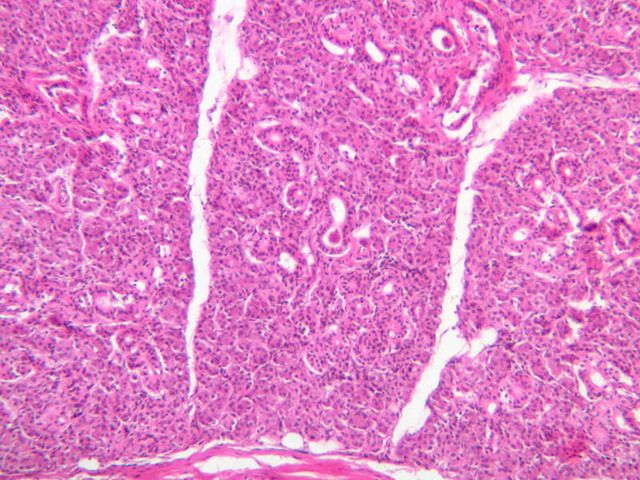
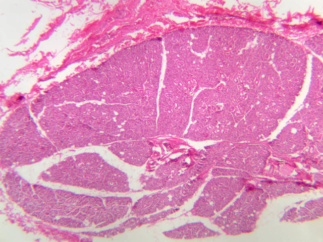
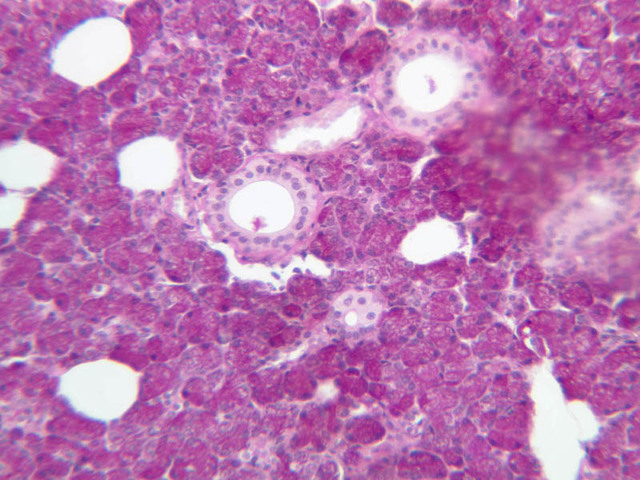
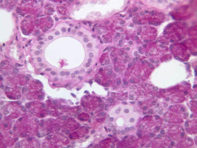
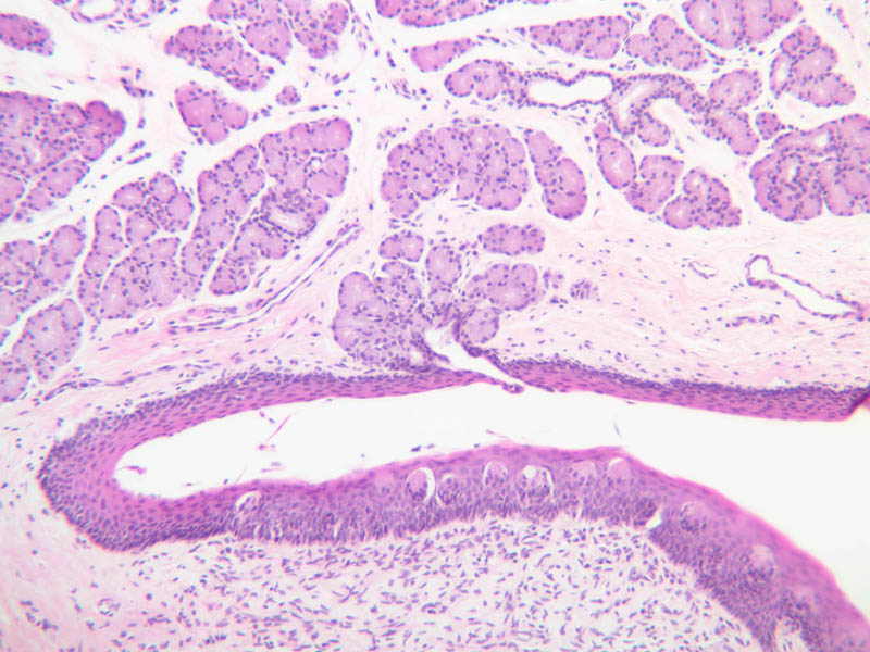
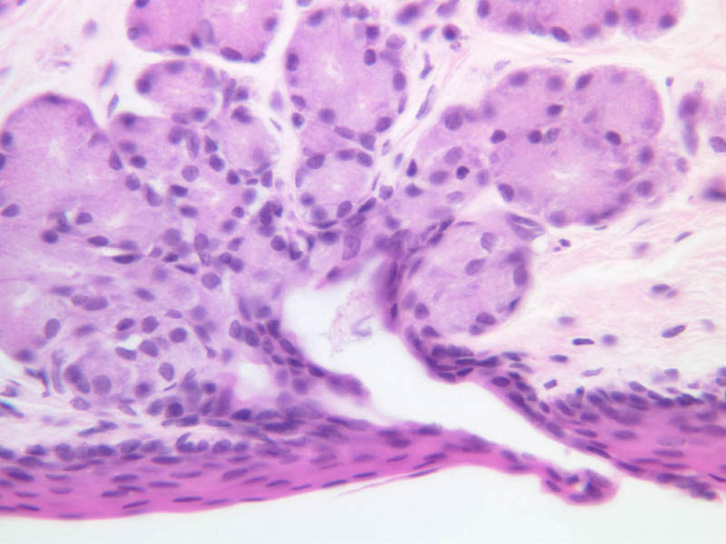
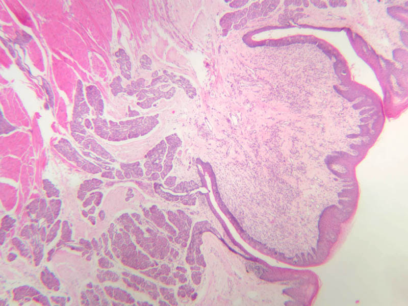
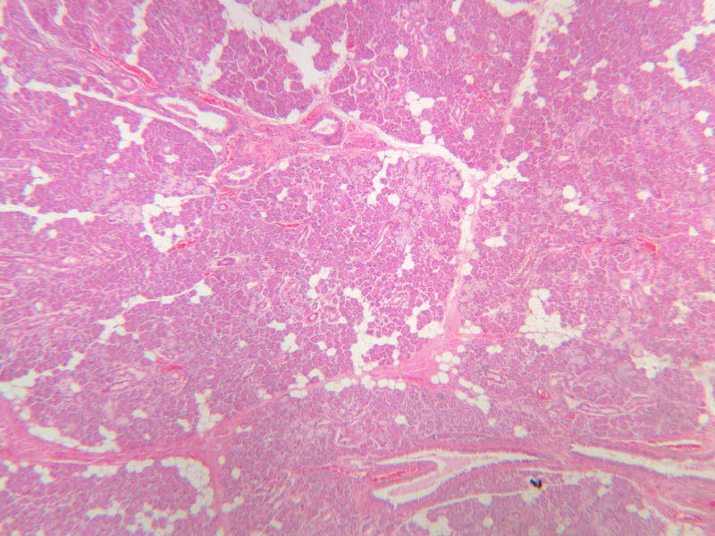
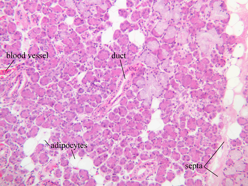
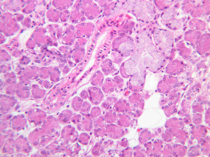
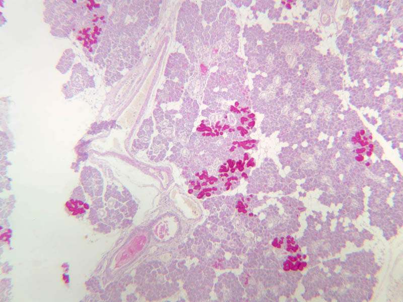
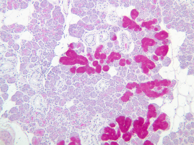
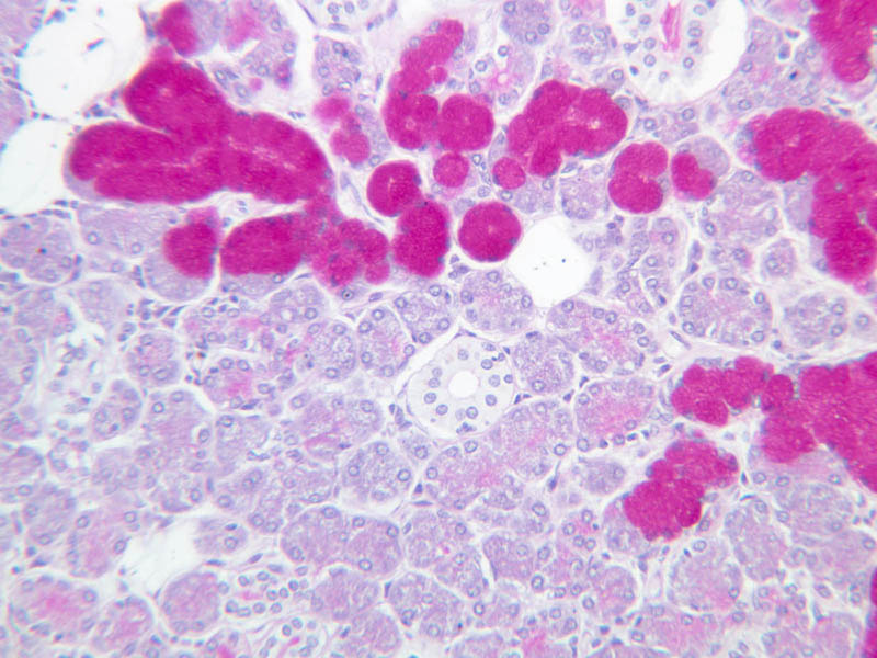
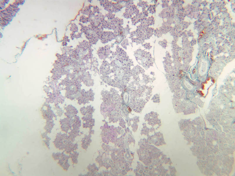
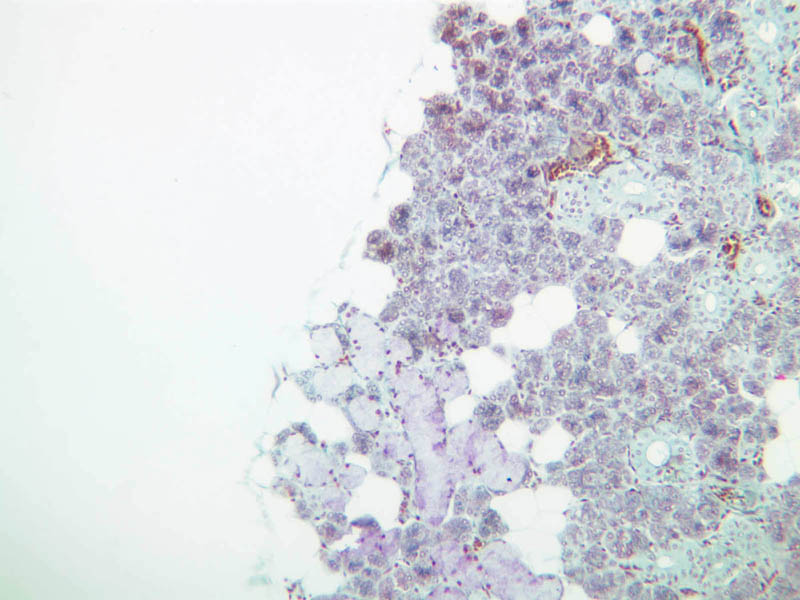
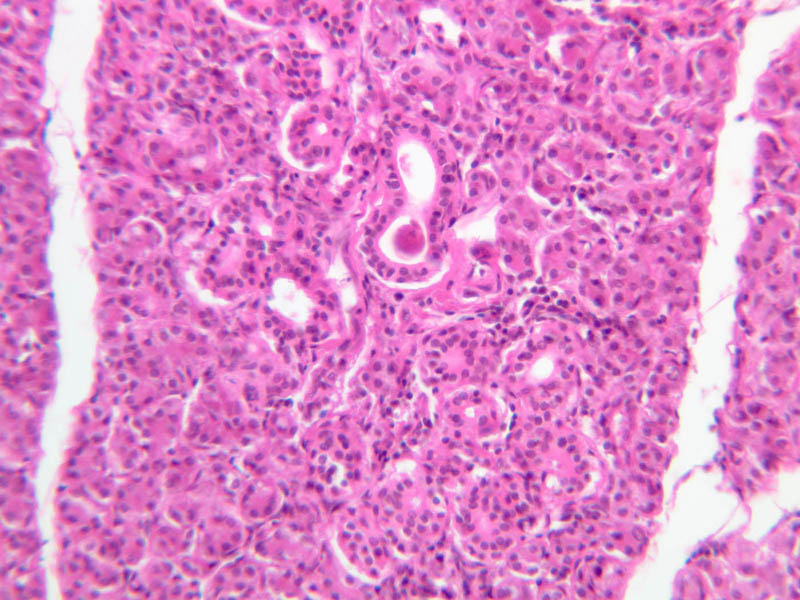
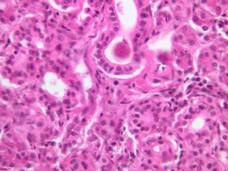
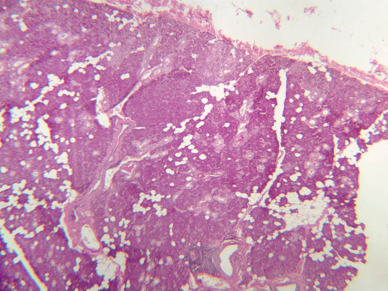
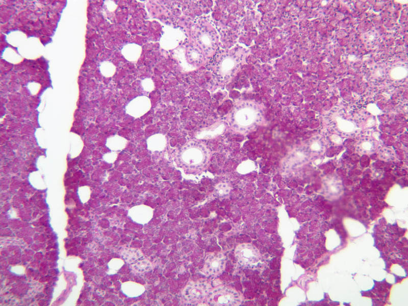
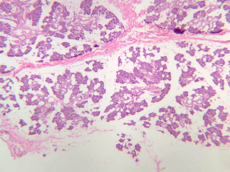
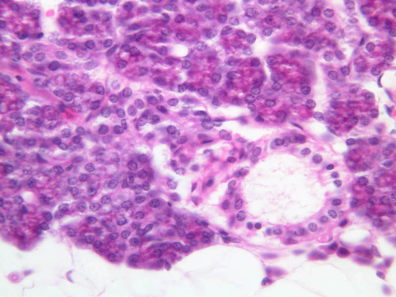
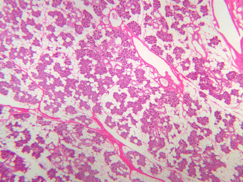
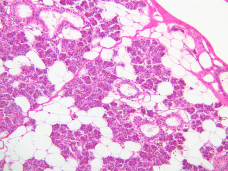
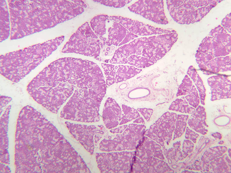
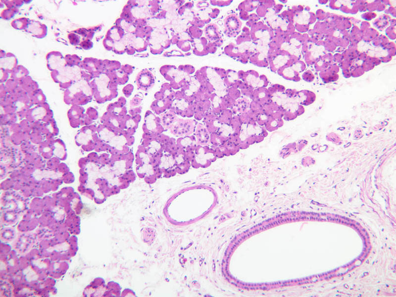
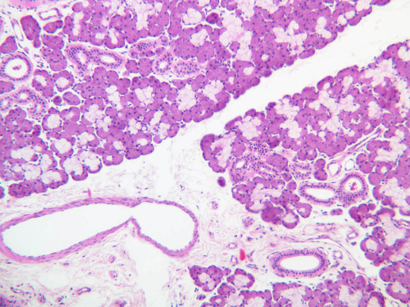
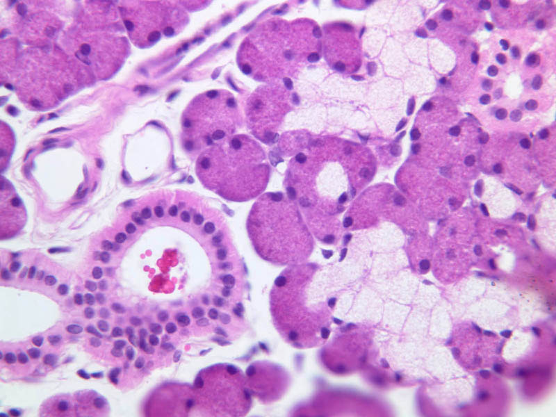
There are three pairs of major salivary glands, demonstrable by dissection as the parotid, submandibular, and sublingual glands. Each of these glands is surrounded by a connective tissue capsule and subdivided into lobes and lobules by connective tissue septa that project from the capsule into the substance of the gland. There are also a large number of small, accessory (or minor) salivary glands, which occur in the submucosa of the lip (labial), palate (palatine), tongue (lingual), cheek (buccal), and gums (gingival or molar). The accessory salivary glands are not encapsulated. There are two types of secretory cells in salivary glands: mucous cells, which produce a viscous secretion consisting largely of highly glycosylated proteins and serous cells, which produce a watery secretion containing the digestive enzymes, amylase and lysozyme. These two cell types occur in different proportions in the different salivary glands. The secretory cells of the parotid gland and the lingual glands of von Ebner are entirely, or almost entirely, serous. Some of the minor salivary glands consist entirely of mucous secreting cells. The submandibular and sublingual glands are obviously mixed seromucous glands, with serous secretory cells predominating in the former, mucous secretory cells, in the latter. Begin your study of salivary glands with the sections of submandibular gland on slide B-43. Each of the three sections on this slide has been stained by a different technique: one section is stained with H&E (B-43 [2.5x, 10x-labeled, 20x, 40x-labeled]), one, by the periodic acid-Schiff (PAS) technique (B-43 [2.5x, 10x, 20x, 40x-labeled]) and one, by the aldehyde fuchsin (AF) technique (B-43 [2.5x, 10x, 20x, 40x]). In the H&E-stained section the zymogen granules of the serous cells are easily discernible, whereas the secretory product of the mucous cells can only be inferred from the vacuolated, unstained appearance of their cytoplasm. By contrast, in the PAS-stained section mucous is rendered a brilliant magenta. In the AF-stained section the apical cytoplasm of mucous cells is pink, while the zymogen granules of the serous cells are a dirty greenish blue. Examine each of these sections to familiarize yourself with the appearance of mucous and serous secretory cells under different staining conditions. Note that serous and mucous cells can be distinguished by strictly morphological criteria--namely, the nuclei of serous cells are round, whereas those of mucous cells are flattened and displaced to the basal portion of the cell. Look for crescent-shaped caps of serous cells (serous demilunes) on some of the mucous acini. These serous cells empty into the acinar lumen by means of canalicules between adjacent mucous cells. Note that mucous secretory units may have either an acinar or a branched tubular morphology. Examine a section of the parotid gland (slides B-46, H & E [2.5x, 10x, 20x, 40x]; B-47, H & E [2.5x, 10x, 20x, 40x]; & B-48, PAS [2.5x, 10x, 20x, 40x]). On low magnification, you can see lobes and lobules of serous acini (or alveoli). Note that the pyramidal cells of the acinus have a spherical, euchromatic nucleus and a markedly granular, well-stained cytoplasm. RER (ergastoplasm) is concentrated in the basal portion and membrane-bound secretory (zymogen) granules in the apical portion of the cells; accordingly, the basal cytoplasm is basophilic and the apical cytoplasm is acidophilic. These features are characteristic of serous cells, of which the human parotid gland is almost exclusively composed. Observe the acinar structure of the parotid gland closely and note that each circlet of epithelial tissue is invested by a basement membrane. Between this basement membrane and the round nuclei of the pyramidal glandular cells themselves, look for the flattened nuclei or eosinophilic processes of myoepithelial cells. Contraction of the processes of these cells expresses the secretory product. The parotid gland contains occasional aggregates of adipocytes, which bear a superficial resemblance to mucous secretory cells. You should be able to persuade yourself that these cells with large unstained vacuoles are not mucous cells by examining the PAS-stained section of parotid gland on slide B-48. Begin your study of salivary gland ducts with the parotid gland (B-46, H & E [2.5x, 10x, 20x, 40x]; B-47, H & E [2.5x, 10x, 20x, 40x]). Note that this gland is divided into lobes and lobules by strands of connective tissue. The branches of connective tissue follow the divisions of the duct system from the oral cavity deep into the gland. As an aid to distinguishing ducts from secretory portions of the gland you should keep in mind that, even at low power, most duct elements are readily apparent because of their eosinophilic cytoplasm. Intercalated ducts, the initial segments of the duct system, are lined by low cuboidal cells and convey the secretory product from acini to striated ducts. Like the intercalated ducts, the striated ducts are intralobular; their simple epithelial lining ranges from cuboidal to high columnar and their eosinophilic cytoplasm is characterized by faint basal striations. These striations are the light microscopic manifestation of deep infoldings of plasma membrane associated with closely packed parallel arrays of mitochondria. This kind of structure is generally thought to subserve active ionic transport. Excretory (interlobular) ducts lie in the connective tissue between lobules. Depending on its caliber, the epithelial lining of an excretory duct may be simple cuboidal or columnar, stratified cuboidal or pseudostratified columnar. In the H & E sections and AF sections of submandibular gland on slide B-44 (H & E [2.5x, 10x, 20x-labeled, 40x], [2.5x, 10x, 20x, 40x]) find examples of ducts of different caliber. You will find that intercalated ducts and striated ducts are evident only in regions with large concentrations of serous acini. This is because mucous secretory portions tend to be of a branched tubular rather than acinar morphology; moreover, ducts that convey mucous secretions serve as simple conduits and do not modify the secretions they convey. The sublingual gland is a mixed seromucous gland with a preponderance of mucous acini. Your slide set does not include a specimen of sublingual gland, but you can get a sense of its architecture by studying the photomicrographs of the parotid and submandibular glands.| Secretory Duct | Location | Diameter | Epithelium | Special Characteristics |
|---|---|---|---|---|
| Intercalated | Intralobular | ~ 5 \xB5m | Low Cuboidal | |
| Striated | Intraloblar | 10-50 \xB5m | Simple Cuboidal | Basal striations |
| Excretory | Interlobular | > 50 \xB5m | Tall cuboidal -> Stratified Columnar | Basal striations |
Salivary Gland Image Gallery


Salivary Gland Table of Identifications
| Row | Structure | Abbreviation | Optimal Stain | Representative Section | Note |
|---|---|---|---|---|---|
| 1 | Blood Vessel | (none) , BV | H&E | |
|
| 2 | Duct | (none) | H&E | |
|
| 3 | Adipocytes | (none), A | H&E, PAS | |
|
| 4 | Septa | (none) | H&E | |
|
| 5 | Serous Acini | SA | H&E, PAS, AF | |
|
| 6 | Mucous Acini | MA | H&E, PAS, AF | |
|
| 7 | Serous Demilune | SD | H&E | |
|
| 8 | Striated Duct | StD | H&E, PAS, AF | |
|
| 9 | Myoepithelial Cells | MC | H&E | |
|
| 10 | Intercalated Duct | ID | H&E, AF | |
|
| 11 | Excretory Duct | ED | H&E | |
|
| 12 | Connective Tissue | CT | AF | |
Top of Page
Epiglottis
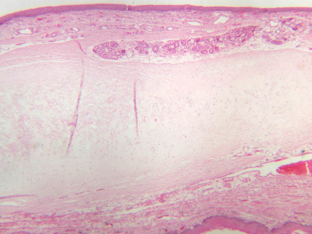
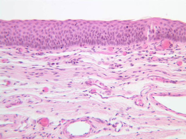
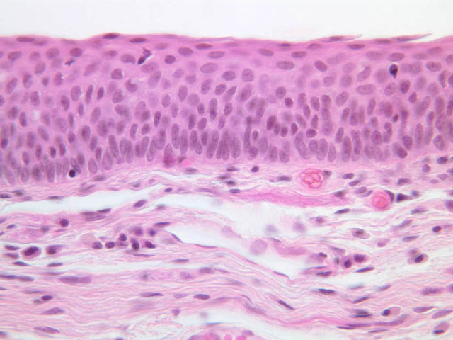
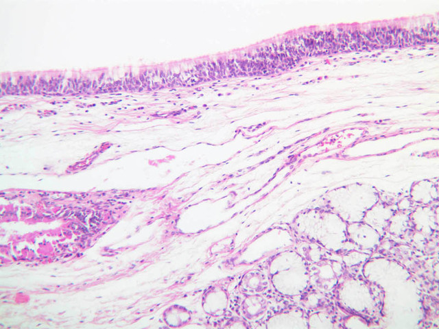
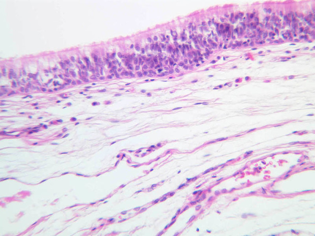
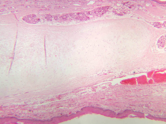
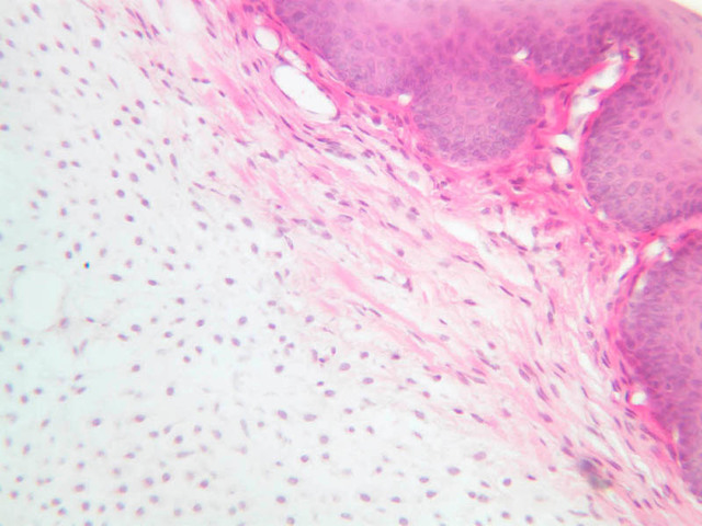
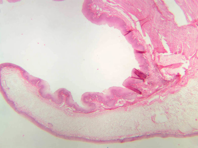
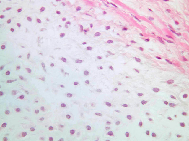
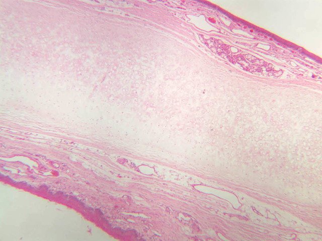
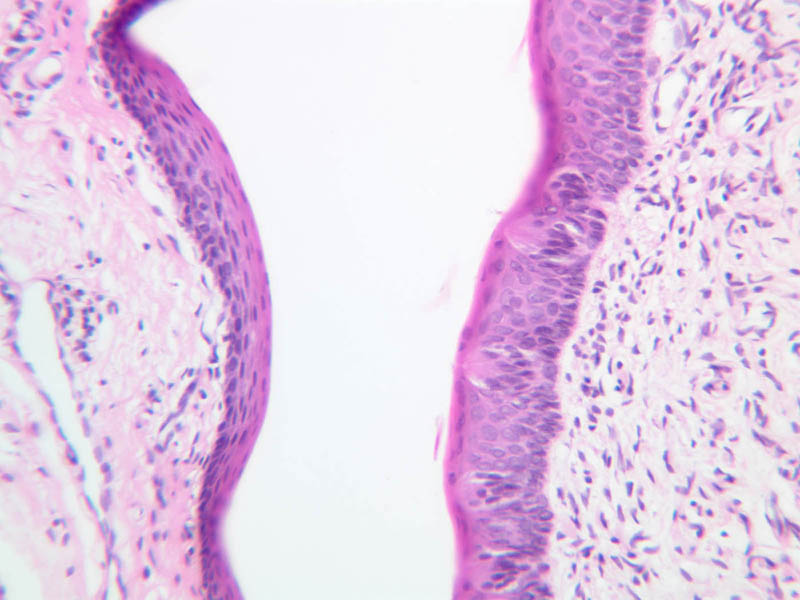
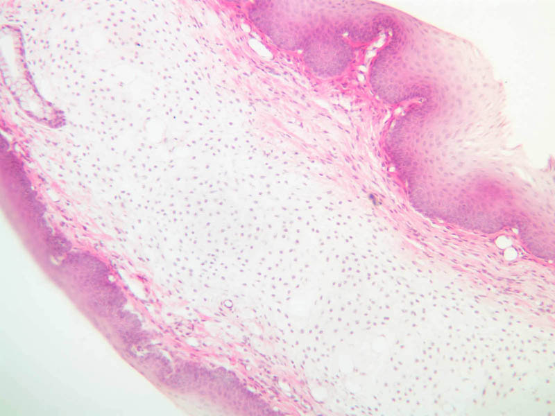
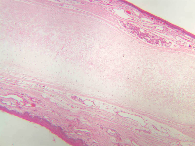
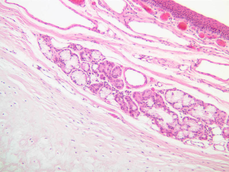
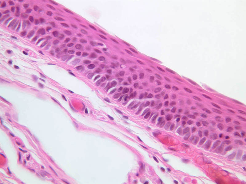
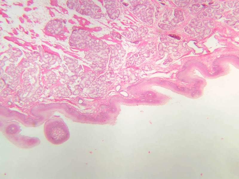
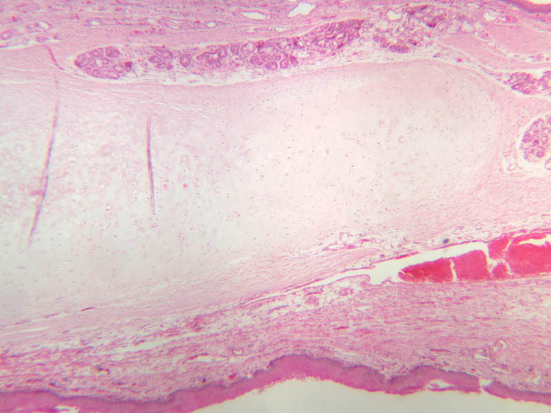
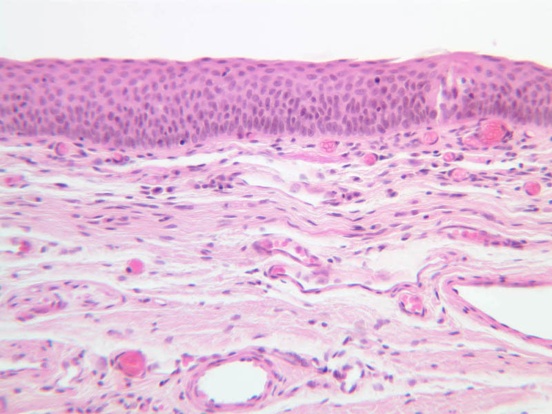
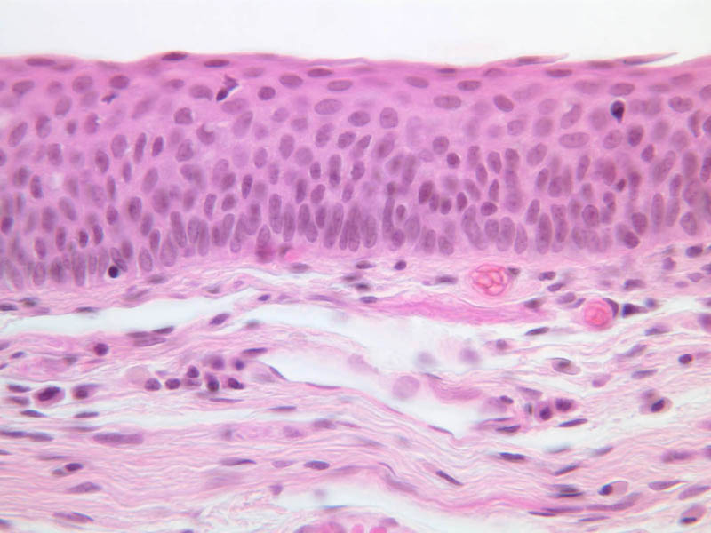
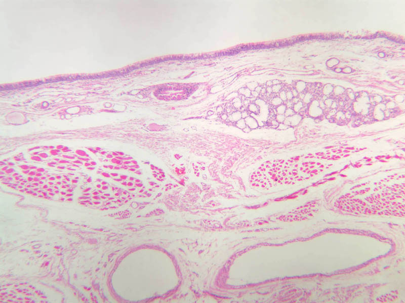
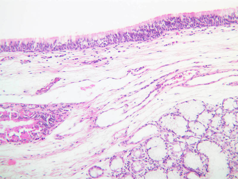
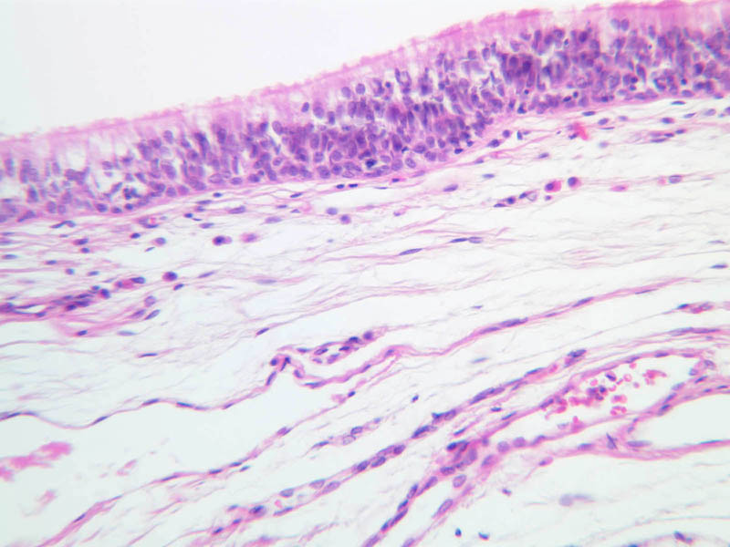
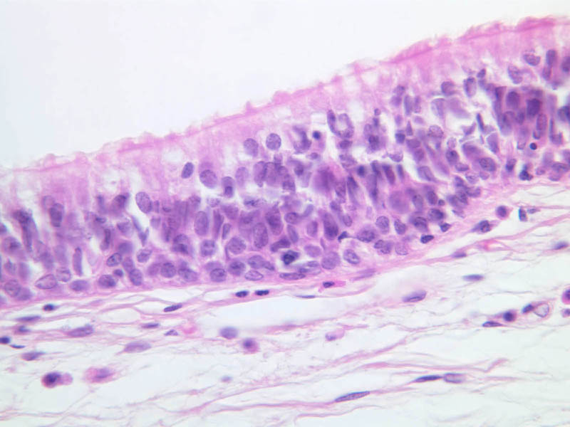
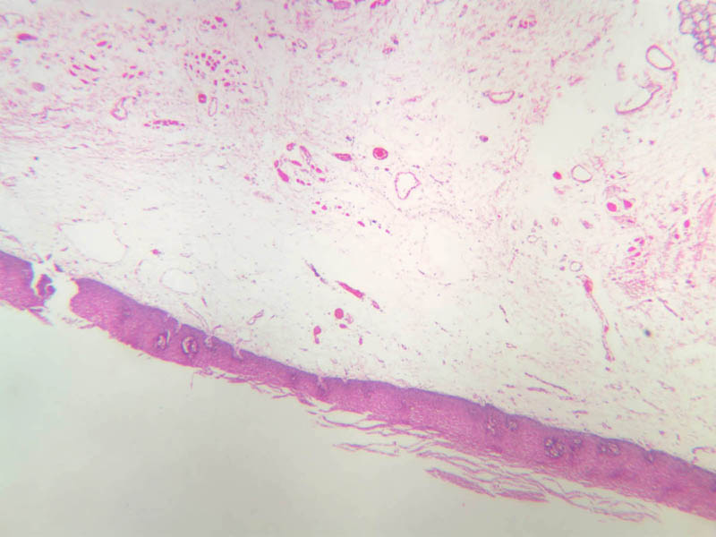
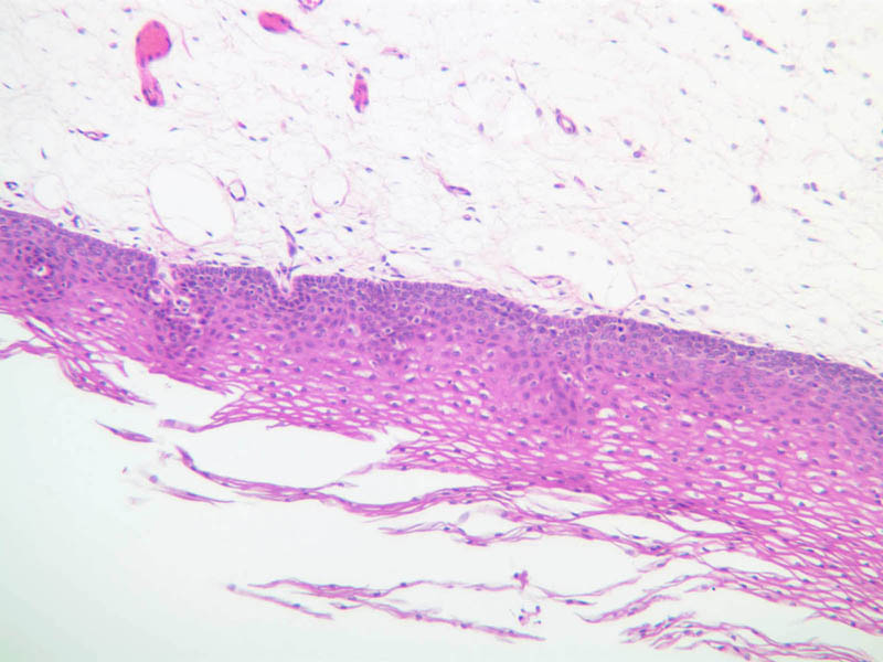
A longitudinal section of the epiglottis appears on slide A-67 (H & E [2.5x, 10x, 20x, 40x], [2.5x, 10x, 20x, 40x]. Find the elastic cartilage forming the core of the epiglottis. (Even though this slide is stained only with H&E, by adjusting the condenser of your microscope you should be able to distinguish the refractile elastic fibers that permeate the cartilage.) You should also be able to find lymphatic tissue and mixed glands in the lamina propria (A-67 [2.5x, 10x, 20x, 40x]). Note that the epithelium on the lingual surface (A-67 [2.5x, 10x, 20x, 40x]) is thicker than that on the pharyngeal surface (A-67 [2.5x, 10x, 20x, 40x]). Why should this be so?Epiglottis Image Gallery


Epiglottis Table of Identifications
| Row | Structure | Abbreviation | Optimal Stain | Representative Section | Note |
|---|---|---|---|---|---|
| 1 | Lingual Surface (of Epiglottis) | (unlabeled) | H&E | |
|
| 2 | Pharyngeal Surface (of Epiglottis) | (unlabeled) | H&E | |
|
| 3 | Seromucous Glands | (unlabeled) | H&E | |
Top of Page
Soft Palate
Examine the section of soft palate on slide A-68 (H & E, Superior Surface [2.5x, 10x, 20x, 40x]; Lingual Surface [2.5x, 10x, 20x, 40x]), noting the elements of connective, muscular and glandular tissue that form its core and the very different epithelial coverage of its superior and inferior surfaces.Soft Palate Image Gallery


Soft Palate Table of Identifications
| Row | Structure | Abbreviation | Optimal Stain | Representative Section | Note |
|---|---|---|---|---|---|
| 1 | Superior Surface (of Soft Palate) | (unlabeled) | H&E | |
|
| 2 | Inferior Surface (of Soft Palate) | (unlabeled) | H&E | |
Top of Page
Chapter Eleven Review
Review of Slides


Review of Identifications
| Row | Structure | Abbreviation | Optimal Stain | Representative Section | Note |
|---|---|---|---|---|---|
| 1 | Vermillion Border | (none) | H&E | |
|
| 2 | Skin (of Lip) | (none) | H&E | |
|
| 3 | Oral Mucosa | (none) | H&E | |
|
| 4 | Keratin | (none) | H&E | |
|
| 5 | Dense Irregular Connective Tissue | (none), CT | H&E, AF | |
|
| 6 | Blood Vessels | (none), BV | H&E | |
|
| 7 | Dorsal Surface (of Tongue) | (none) | H&E | |
|
| 8 | Ventral Surface (of Tongue) | (none) | H&E | |
|
| 9 | Skeletal (Striated) Muscle | (none), M | H&E | |
|
| 10 | Filiform Papillae | (none) | H&E | |
|
| 11 | Circumvallate Papilla | (none) | H&E | |
|
| 12 | Cleft | (none) | H&E | |
|
| 13 | Taste Bud | TB, (circled areas) | H&E | |
|
| 12 | Serous (von Ebner's) Glands | Gl | H&E | |
|
| 13 | Duct (tongue) | D | H&E | |
|
| 14 | Stratified Squamous Epithelium | SSE | H&E | |
|
| 15 | Fungiform Papillae | (none) | H&E | |
|
| 16 | Serous (von Ebner's) Glands | Gl | H&E | |
|
| 17 | Foliate Papillae | (none) | H&E | |
|
| 18 | Duct (gland) | (none) | H&E | |
|
| 19 | Adipocytes | (none), A | H&E, PAS | |
|
| 20 | Septa | (none) | H&E | |
|
| 21 | Serous Acini | SA | H&E, PAS, AF | |
|
| 22 | Mucous Acini | MA | H&E, PAS, AF | |
|
| 23 | Serous Demilune | SD | H&E | |
|
| 24 | Striated Duct | StD | H&E, PAS, AF | |
|
| 25 | Myoepithelial Cells | MC | H&E | |
|
| 26 | Intercalated Duct | ID | H&E, AF | |
|
| 27 | Excretory Duct | ED | H&E | |
|
| 28 | Lingual Surface (of Epiglottis) | (unlabeled) | H&E | |
|
| 29 | Pharyngeal Surface (of Epiglottis) | (unlabeled) | H&E | |
|
| 30 | Seromucous Glands | (unlabeled) | H&E | |
|
| 31 | Superior Surface (of Soft Palate) | (unlabeled) | H&E | |
|
| 32 | Inferior Surface (of Soft Palate) | (unlabeled) | H&E | |
Top of Page
Comments
Top of page -- AshleyLPistorio - 27 May 2007Edit | Attach | Print version | History: r2 < r1 | Backlinks | View wiki text | More topic actions
Topic revision: r2 - 20 Jun 2015, LorenEvey

- Epithelium
- Connective Tissue
- Muscle
- Nervous Tissue
- Cardiovascular System
- Skin Appendages and Sensory Receptors
- Lymphatic System
- Cartilage and Bone
- Respiratory System
- Peripheral Blood and Bone Marrow
- Oral Cavity and Salivary Glands
- Esophagus and Gastrointestinal Tract
- Pancreas, Liver, and Gall Bladder
- Endocrine Organs
- Male Reproductive System
- Female Reproductive System
- Urinary System
Ideas, requests, problems regarding Medical Histology? Send feedback